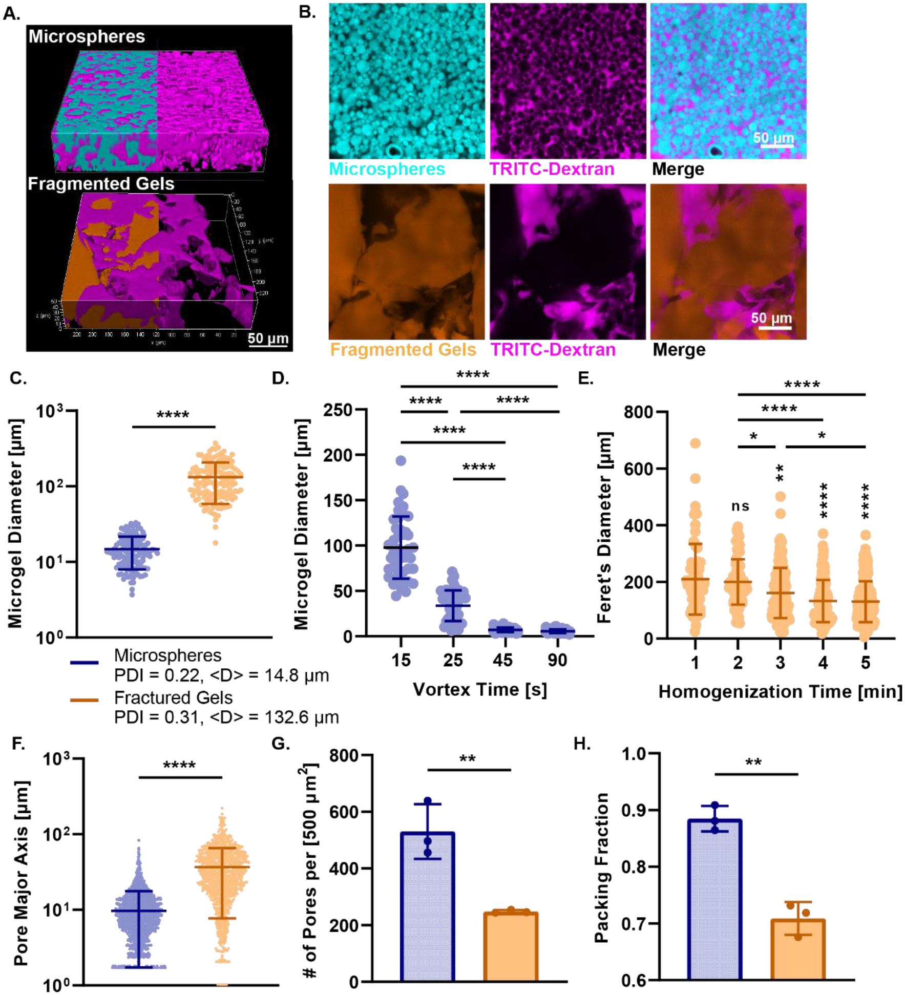
Figure 2.

Particle and Pore Size Analysis of Granular Hydrogels. (A) 3D rendered confocal images of Alexa Fluor-MAL-488 labeled microspheres (cyan) and fractured gels (orange) incubated with high molecular weight TRITC-dextran (magenta). (B) Confocal images of microspheres and fragmented gels with TRITC-dextran infiltration and merge. (C) Microgel diameter for microspheres and fractured gels. Microgel diameter of fractured gels is greater than microspheres and the polydispersity index is much higher for fractured gels. (D) Demonstration of the control over size of microsphere through alteration of vortex time from 15 seconds to 90 seconds. (E) Lack of control over size of fragmented gels through mechanical fragmentation from 1 minute homogenization to 5 minutes. Significance from 1 min time group displayed above groups, all other comparisons are displayed as bars above all groups. (F) Interparticle distance between pores of scaffold for microspheres and fractured gels as measured by the pore major axis of each pore. (G) The number of pores per area for both microspheres and fragmented gels. (H) The packing fraction of microspheres and fragmented gels. For all panels, significance between means of three or more groups determined by one-way ANOVA, p < 0.05 with Tukey’s post-hoc pairwise comparisons, significance between means of two groups determined by two-tailed student’s t-test. * = p < 0.05, ** = p < 0.01, *** = p < 0.001, **** = p < 0.0001.
