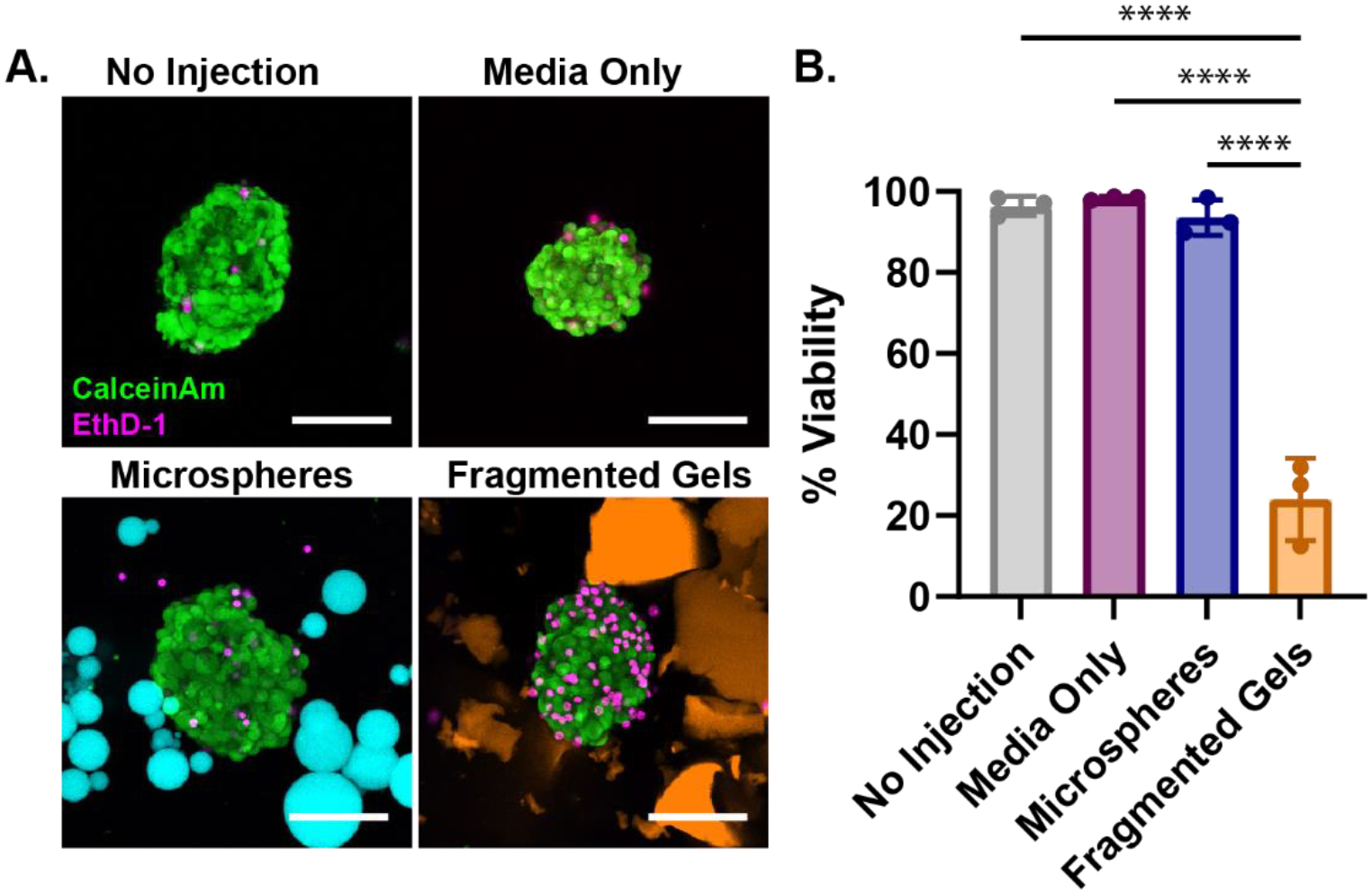
Figure 6.

Injection of Islets within Granular Hydrogels. (A) Human pancreatic islets injected in microspheres (cyan), fragmented gels (orange), media only as compared to a no-injection control. Islets were stained with Calcein AM (Live, green) and EthD-1 (dead, red) after injection, PEG-MAL gels are labeled with AlexaFluor 647. Media was added to dilute and disperse the microspheres and fragmented gels after injection to aid in identifying and imaging islets. Scale bar is 50 μm. (B) Percent viability of the islets after injection within media-only, microspheres, fragmented gels, as compared to a no-injection control. Percent viability was calculated by dividing the number of live cells by the total number of cells in the islet, data is representative of multiple donors. Significance between means of three or more groups determined by one-way ANOVA, p < 0.05 with Tukey’s post-hoc pairwise comparisons. * = p < 0.05, ** = p < 0.01, *** = p < 0.001, **** = p < 0.0001.
