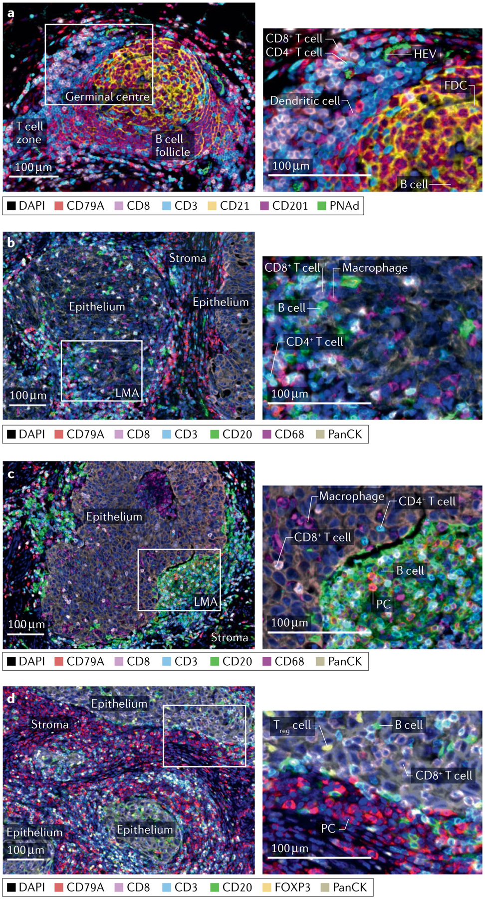
Fig. 3 |. TIL-B neighbourhoods.

a–d | Multiplex immunofluorescence staining of untreated human high-grade serous ovarian cancer (parts a–c) and medullary breast cancer tissues (part d) showing examples of a secondary follicular tertiary lymphoid structure (part a), astromal lympho-myeloid aggregate (LMA) with infiltration of a neighbouring epithelial region by CD4 and CD8 T cells and B cells (part b), a dense infiltrate of B cells restricted to the stromal compartment despite infiltration of adjacent epithelium by T cells and macrophages (part c) and a dense stromal infiltrate of plasma cells (PCs) accompanied by intra-epithelial B cells (part d). Left column: low-magnification views with relevant structural zones labelled. Right column: high-magnification views of boxed regions from left column showing examples of CD8 T cell (CD8+CD3+), presumptive CD4 T cell (CD8−CD3+), dendritic cell (CD201+), follicular dendritic cell (FDC; CD21+), high endothelial venule (HEV; PNAd+), B cell (CD20+), PC (CD20−CD79A+), macrophage (CD68+) and regulatory T cells (Treg cells; CD3+FOXP3+). Markers and associated colours specified below each image. Epithelium, tumour epithelium; FOXP3, forkhead box protein P3; PanCK, pan cytokeratin; PNAd, peripheral node addressin; stroma, tumour stroma; TIL-B, tumour-infiltrating B lymphocyte. Image courtesy of Katy Milne, Tashi Rastogi and Karanvir Singh, BC Cancer.
