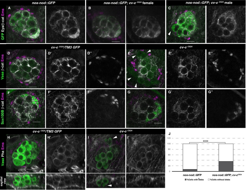
Figure 3. Gonad morphology in cv-c mutant embryos.
(A–C) Gonads with germ cells labelled with nos-nod::GFP (green), pigment cells with anti-Ems (magenta), and male-specific somatic gonad precursor (msSGP) with anti-Eya (nuclear grey staining) and the AJs with anti-β-catenin (grey membranes). Right panels show Eya and β-catenin channel. (A) In the control testis, germ cells are ensheathed by thin mesoderm extensions produced by the interstitial cells detectable by β-catenin staining. Similar germ cell ensheathment is observed in cv-cC524 ovaries (B), while in cv-cC524 testis (C) some germ cells become extruded from the gonad and are not enveloped by β-catenin (arrowhead). (D–G) Testes labelled with mesodermal specific markers to detect the pigment cells (Ems, magenta D–G) or the msSGPs (Sox100B, green F, G) in heterozygous (D, F) or cv-cC524 homozygous mutant embryos (E, G). Grey channels in right panels correspond to β-catenin in (D-G), Ems in (D, E), or Sox100B in (F, G). All mesoderm cell types are specified in cv-c mutant testis despite morphological aberrations resulting in the pigment cell layer’s discontinuity (compare D and F with E and G). (H–J) Testes stained with anti-Vasa (green) to label the germ cells and phalloidin (grey and right panels) to show actin filaments in heterozygous (H) or homozygous cv-cC524 mutants (I). Germ cells in mutant testes present protrusions compatible with migratory movements (arrowheads). Z sections are shown below H, I panels. Scale bar: 10 µm. (J) Quantification of blebbing cells in wild-type or mutant cv-c background using Fisher test; ****p value <2.2e−16 (nos::GFP N = 575 and nos::GFP;cv-cC524 N = 744) (Figure 3—source data 1).


