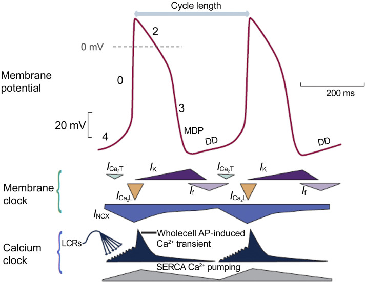
Figure 3.
Schematic of sinus node membrane potential and underlying mechanisms. Top: action potential (AP) of a rabbit sinoatrial nodal myocyte (red). Middle: different components of the “membrane clock.” Bottom: different components of the “Ca2+ clock.” During phase 4, cytosolic [Ca2+] increases due to spontaneous Ca2+ releases from the sarcoplasmic reticulum (SR) (dark blue). Activation of L-type Ca2+ channels (orange) then causes Ca2+-induced Ca2+ release from the SR, resulting in the whole cell Ca2+ transient. Cytoplasmic Ca2+ is removed by the sarco(endo)plasmic reticulum calcium ATPase (SERCA) pump (gray) and by sodium-calcium exchanger (NCX) (blue). DD, diastolic depolarization; ICa,T, T-type voltage-dependent Ca2+ current; ICa2,L, L-type voltage-dependent Ca2+ current; If, funny current; INCX, sodium-calcium exchange current; IK, delayed rectifier potassium current; LCRs, local Ca2+ releases; MDP, maximum diastolic potential. Reproduced from Ref. 79 with permission.

