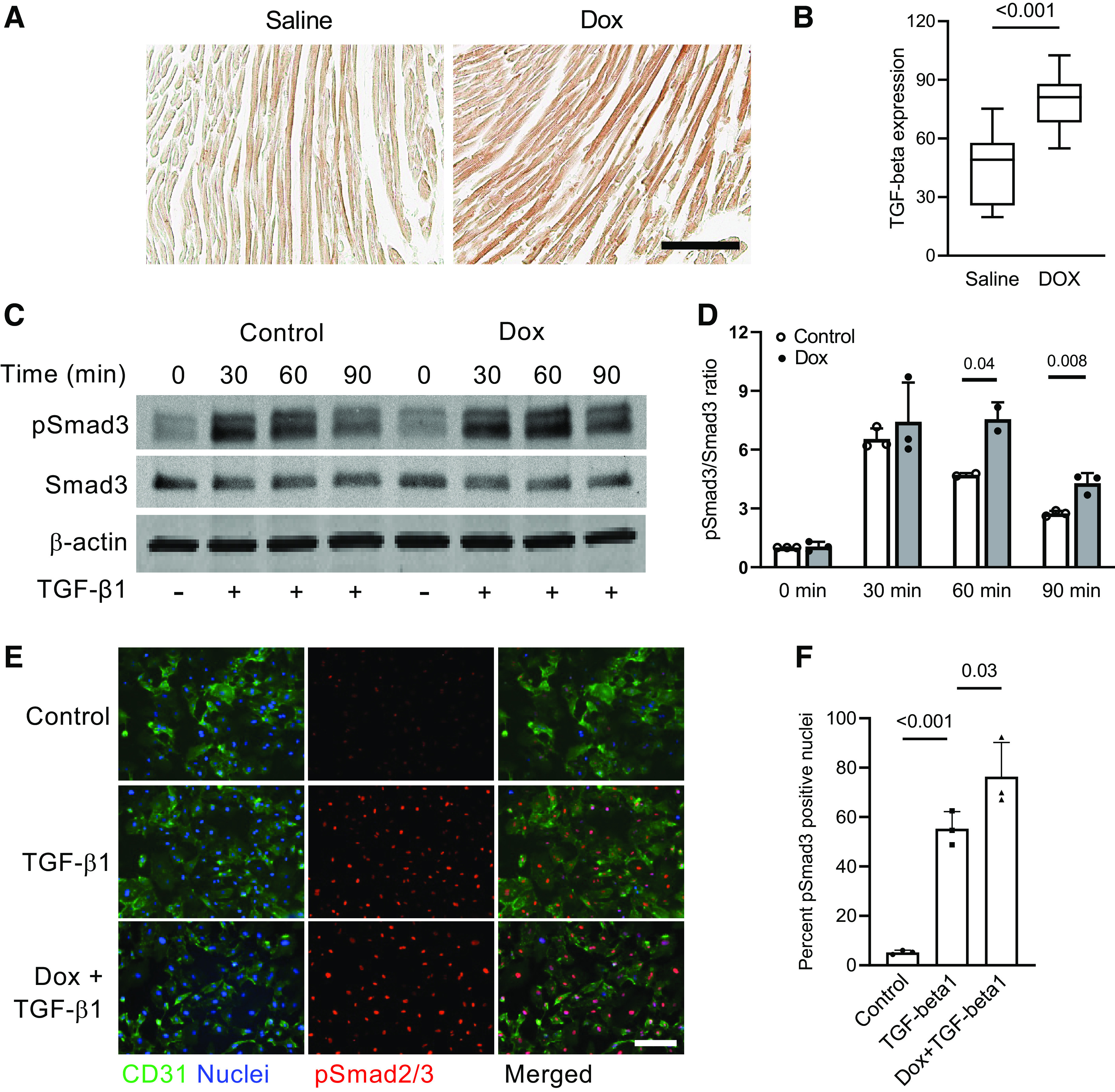
Figure 2.

Doxorubicin enhances activity of the canonical TGF-β pathway in endothelial cells. A: mice were intravenously injected with doxorubicin (5 mg/kg) or saline, and heart tissues were harvested 24 h after the injections and processed for immunohistochemical staining. Cardiac sections were stained with the TGF-β pan-specific antibody and analyzed using color bright-field microscopy. Scale bar = 100 µm. B: TGF-β expression in cardiac tissues was measured by density of horseradish peroxidase substrate 3,3′-diaminobenzidine (DAB) deposits. n = 3 hearts per group. Eight to ten randomly selected fields per heart were analyzed. P value for the unpaired two-tailed t test is shown. C: human cardiac microvascular endothelial cells were pretreated with 16 nM doxorubicin (Dox) for 72 h, starved for 4 h, and incubated with 0.3 ng/mL TGF-β1 for 0 to 90 min. Protein extracts were then processed according to the immunoblotting protocol and incubated with the indicated antibodies. pSmad3, phospho-Smad3. D: pSmad3 band intensity in cells lysed at the indicated time points after treatment with TGF-β1 is presented as a ratio of phospho- to total Smad3. Results of 3 independent experiments are shown. P values for the unpaired two-tailed t tests are shown. E: increased pSmad2/3 nuclear translocation response to TGF-β1 in endothelial cells pretreated with 16 nM Dox for 72 h, starved for 4 h, and treated with 0.3 ng/mL TGF-β1 for 1 h. Endothelial cells were labeled using CD31 antibody, and nuclei were stained with Hoechst 33342. Scale bar = 200 µm. F: quantification of pSmad2/3-positive nuclei (as percentage of total nuclei) in endothelial cells treated with Dox and/or TGF-β1. Results of 3 independent experiments are shown. P values for the one-way ANOVA tests are shown.
