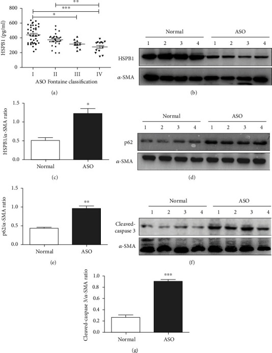
Figure 1.

Expression of HSPB1 in ASO patients. (a) ELISA was used to quantify the plasma levels of HSPB1. The absorbance was measured using a microplate spectrophotometer at 450 nm with wavelength correction set to 630 nm. The error bars are the SDs of 3 replicates. ∗p < 0.05, ∗∗p < 0.01, and ∗∗∗p < 0.001. (b–g) Sclerotic intima was collected from four patients clinically diagnosed with grade III ASO, and normal intima obtained from amputation patients was used as a control. The tissues were homogenized in RIPA buffer containing complete protease inhibitor, and then, Western blot analysis was performed to measure the protein expression of (b) HSPB1, (d) p62, and (f) cleaved caspase-3. Densitometry was used to determine the fold change in the expression of (c) HSPB1, (e) p62, and (g) cleaved caspase-3 relative to the expression of β-actin (n = 6 for each group; ∗p < 0.05, ∗∗p < 0.01, and ∗∗∗p < 0.001).
