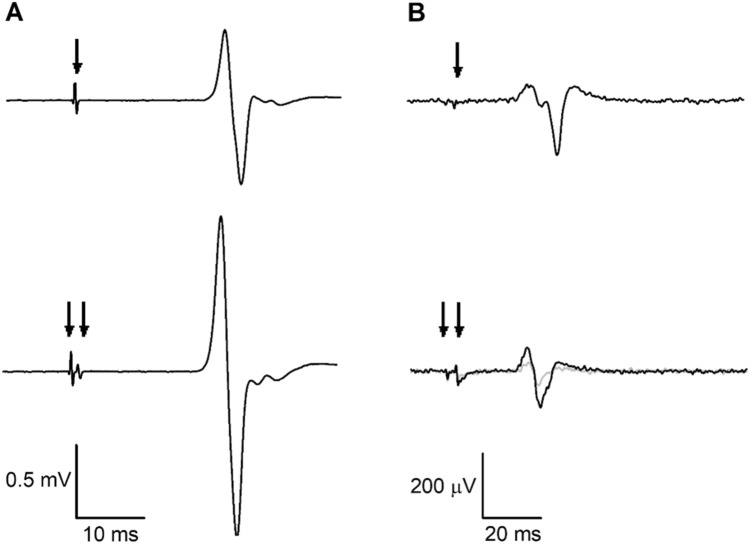
Fig. 2.
Representative electromyographic (EMG) traces showing motor-evoked potentials (MEPs) from the nondominant first dorsal interosseous (FDI) muscle of a single participant. Arrows indicate transcranial magnetic stimulation (TMS) artefacts. A Short-interval intracortical facilitation (SICF). Top trace: non-conditioned MEP with suprathreshold first stimulus (S1) given alone; bottom trace: conditioned MEP with S1 followed by subthreshold second stimulus (S2). B Short-interval intracortical inhibition (SICI). Top trace: non-conditioned MEP with test stimulus given alone (threshold-hunting target, THT; 200 µV); bottom trace: conditioned MEP, where the grey trace represents unadjusted THT and the black trace represents adjusted THT in the presence of the conditioning stimulus

