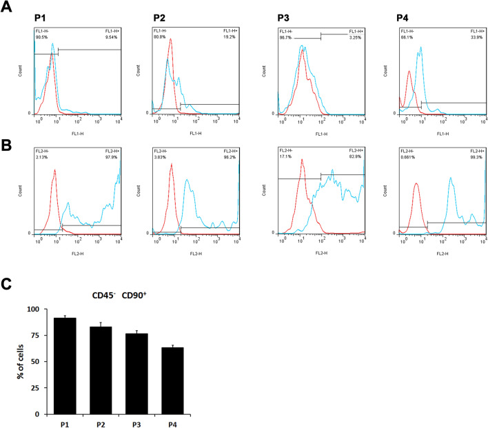
Fig. 1.
Characterization of adipose tissue-isolated MSCs (ADSCs). Primary rat MSCs isolated from adipose tissue were stained with IgG control (red lines) or A the FITC-conjugated anti-rat CD45 monoclonal antibody (blue lines) and B the PE-conjugated anti-rat CD90 monoclonal antibody (blue lines) throughout ADSC passages 1–4 (P1-P4. The freshly isolated ADSC is passage 0, P0). The black lines indicated the dividing boundary of negative (IgG control) and positive staining signals. C The percentage of CD90 positive and CD45 negative cells in different primary ADSC culture passages. In passage 1 isolated cells, the percentage of ADSCs were the highest among the first four passages

