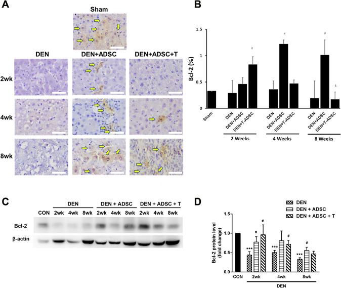
Fig. 8.
Bcl-2 levels were decreased in DEN-injected liver tissues and ADSCs treatments increased Bcl-2 levels. A Immunohistochemical Bcl-2 staining in experimental animal liver tissues with DEN treatment for 2, 4, or 8 weeks (DEN), respectively (2wk, 4wk, 8wk). DEN+ADSC: DEN-injected liver tissue slices treated with ADSC for another 4 weeks. DEN+ADSC+T: DEN-injected liver tissue slices with ADSCs pretreated with L-theanine. Arrow point to the brown positive Bcl-2 signal stain. The scale bar represents 25 μm. B Quantification of immunohistochemical Bcl-2 staining in experimental animal liver tissues. C Western blotting analysis and D quantification of Bcl-2 expression in experimental animal liver tissues. Compared to Sham, ***: p < 0.001. Compared to DEN, #: p < 0.05. Compared to DEN + ADSC, $: p < 0.05

