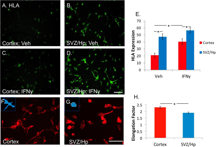
FIGURE 3.
Microglia from the ventricular/Hp region express greater levels of HLA-DP, DQ, and DR are more rounded than cortical microglia. (A) In basal conditions without any treatment, a variable level of HLA-DP, DQ, DR is expressed by cortical adult human microglia. (B) Microglia from ventricular/Hp regions have a higher basal level of HLA-DP, DQ, DR expression. (C) IFNy (1 ng/ml, 96 h) increased cortical microglial expression of HLA-DP, DQ, DR, as well as that of ventricular/Hp microglia (D). Scale bar = 100 μm. (E) Quantification of HLA expression (arbitrary fluorescence units) showing differential expression by cortical and ventricular/Hp microglia, and a significant increase in HLA expression with IFNy treatment. (F) Adult human microglia isolated from cortical tissue immunolabelled with the cell surface marker CD45 have a heterogeneous morphology with various extended processes. (G) Microglia isolated from ventricular/Hp regions have a rounder morphology with fewer processes. Insets, in panels (F,G) show representative morphology of cells. Scale bar = 100 μm. (H) Quantification of microglial morphology using Metamorph Elliptical Form Factor (a measure of elongation) image analysis demonstrates a significant difference in microglia shape between cortical and adjacent neurogenic regions. *indicates P value < 0.05.

