Figure 1.
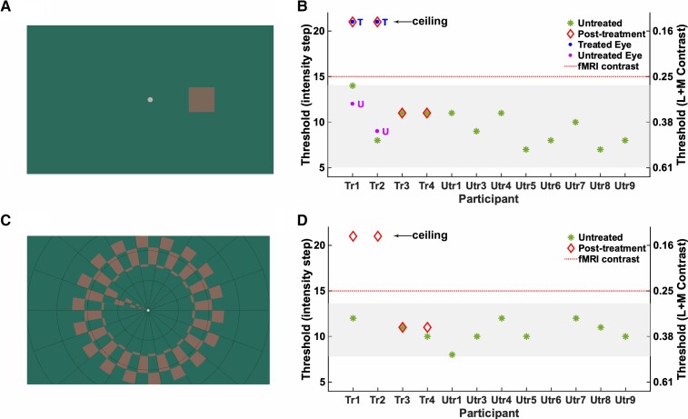
Cone selective stimuli and psychophysics. (A) Example of the lowest cone-selective contrast tested psychophysically, embedded in the 4AFC square localization psychophysics task. Participants judged the position of a target (size 3°), presented 6° to the left, right, above or below centre, with unlimited time to search and uncontrolled gaze. Note that stimulus appearance is screen-dependent. (B) Binocular contrast discrimination thresholds in the 4AFC square localization psychophysics task (50% correct) for children with ACHM. Left y-axis indicates contrast detection thresholds in units of staircase step with decreasing stimulus intensity (1 = highest contrast, 21 = lowest contrast), with the right y-axis indicating the corresponding L + M cone Michelson contrasts (see ‘Validating cone selective stimuli' in the Supplementary material for details). Green stars indicate measures for 12 untreated patients with ACHM. Shaded area: 95% prediction interval computed using the t-distribution quantile. Follow-up measures 6–14 months after treatment are shown for treated Patients Tr1–Tr4 (red diamonds). Post-treatment measures for Patients Tr1 and Tr2 were repeated monocularly for the treated eye (blue dots, ‘T’) and untreated eye (purple dot, ‘U’). (C) Cone-selective pRF mapping stimulus presented inside the scanner (at fixed contrast indicated by red dotted line in B and D), and in the Ridge motion discrimination task (maximum eccentricity 8.6°). In the scanner, participants detected target dimming at fixation. In the Ridge task they discriminated direction of ring movement (inward/outward). (D) As in B but showing binocular contrast discrimination thresholds (at 50% correct) for the Ridge motion discrimination task performed with the pRF mapping stimulus.

