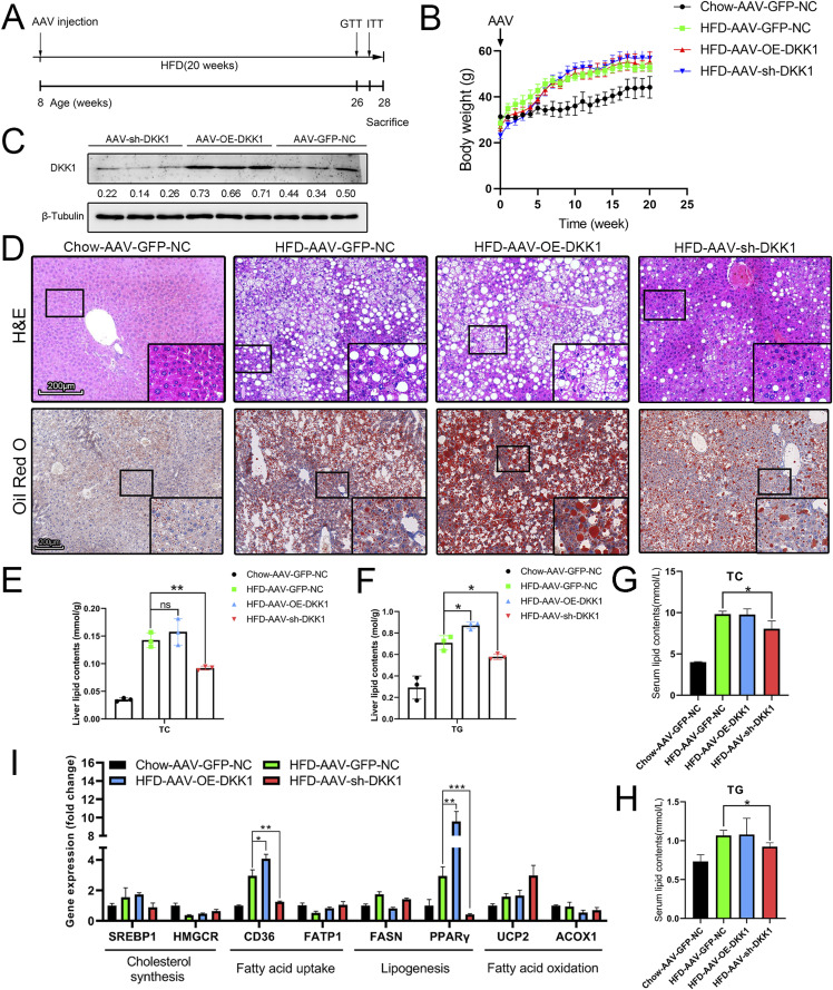
Figure 2. Liver-specific DKK1 overexpression exacerbates HFD-induced liver steatosis.
(A) Schematic illustration of experiment procedure. AAV tail intravenous injection with AAV-GFP-NC (n = 4), AAV-OE-DKK1 (n = 5), or AAV-sh-DKK1 (n = 6) and then fed HFD for 20 wk before euthanasia. (B) Dynamic body weight tracking of chow- and HFD-fed mice with DKK1 manipulations. (C) Representative Western blot of DKK1 in liver samples of HFD-fed mice after 20 wk (n = 3). Each lane represents liver lysates from individual mouse. (D) H&E and ORO staining. Scale bar = 200 µm; (E, F) lipid contents in liver of mice with different DKK1 gene manipulations under chow or HFD fed (n = 3). (G, H) Serum lipid contents with different DKK1 gene manipulations under chow or HFD fed. (I) The expression confirmations of lipid metabolism–related genes in liver of mice with different DKK1 gene manipulations under chow or HFD fed (n = 3). *P < 0.05, **P < 0.01, ***P < 0.001 as compared with the indicated controls by two-tailed t tests. All data are shown as the means ± SD. ns, not significant.
Source data are available for this figure.

