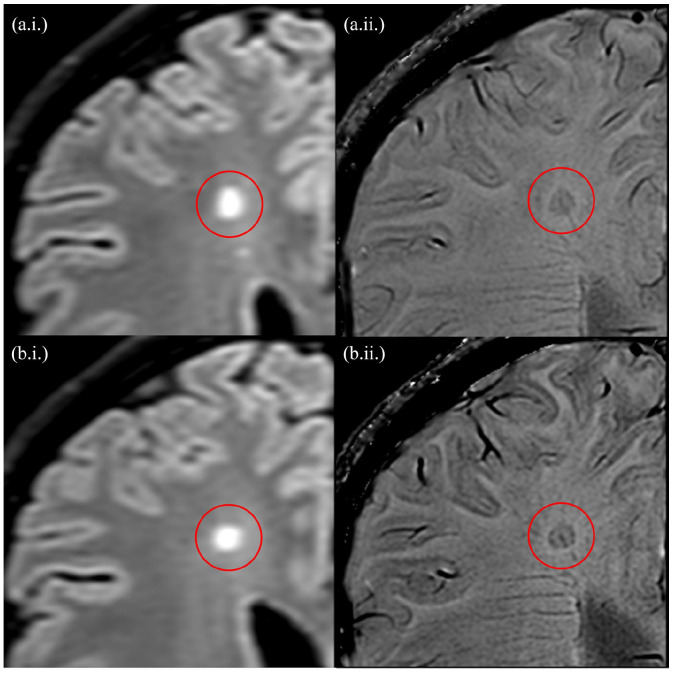
Figure 1.
Consecutive slices of a paramagnetic rim lesion (with a central vein) detected using the fluid-attenuated inversion recovery (a.i. and b.i.) and phase-sensitive imaging (a.ii. and b.ii.), at 3T. As per the study protocol, the lesions demonstrate a hypointense, ring-like structure corresponding to the lesion edge which is present on at least two consecutive slices.

