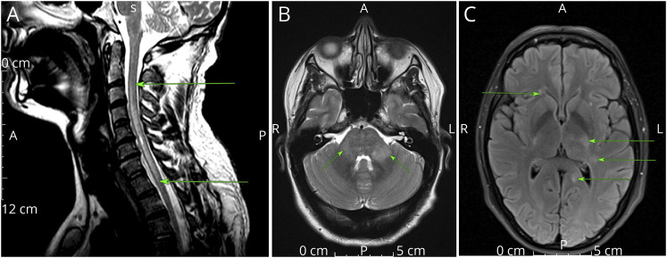
Figure 1. MOGIgG+ MRI.
(A) T2-weighted cervical spinal cord MRI. LETM (arrows), typical of MOGAD. Patient also tested positive for Borrelia burgdorferi. (B) Axial T2-weighted MRI. Bilateral asymmetric T2 hyperintensities in the middle cerebellar peduncles (arrows), typical of cerebral MOGAD. (C) Axial FLAIR MRI. Multiple hyperintensities involving left trigone, posterior limb of left internal capsule, and frontal horn right lateral ventricle (arrows). Abbreviations: LETM = longitudinally extensive TM; MOGAD = myelin oligodendrocyte glycoprotein antibody–associated disease

