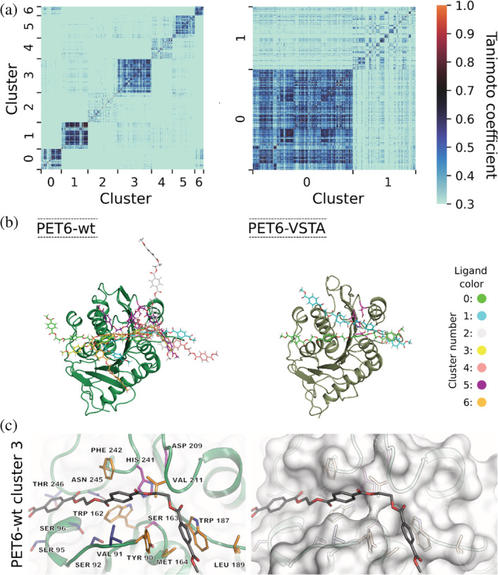
FIGURE 5.

Binding modes of a PET tetramer in complex with PET6‐wt and PET6‐VSTA. (a) Tanimoto similarity matrices clustered according to similar interaction patterns representing clusters of different binding modes. (b) Superimposition of representative structures of the identified binding modes corresponding to the clusters in (a). (c) Detailed representation of residues frequently involved in enzyme–ligand interaction for PET6‐wt. The left side shows the enzyme in cartoon representation with the interacting residues as well as the catalytic triad (Ser163, Asp209, and His241) depicted in sticks. The residues colored in orange are involved in interactions in both PET6‐wt and PET6‐VSTA, while those shown in blue are additionally relevant in the context of PET6‐VSTA; the catalytic triad is marked in purple. The same perspective is shown on the right but with a less transparent surface to visualize the positioning of the ligand within the binding region
