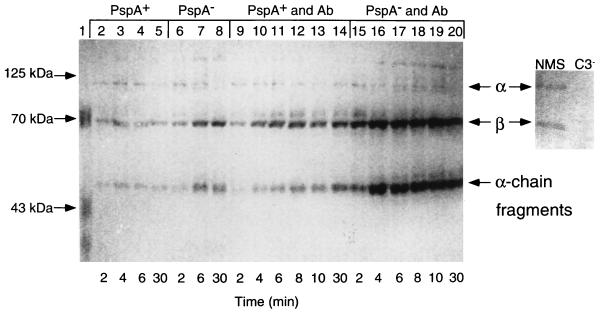
FIG. 3.
Western blot analysis of C3 deposition onto PspA+ and PspA− pneumococci. Pneumococci were exposed to XID mouse serum in the absence (lanes 2 to 8) or presence (lanes 9 to 20) of anticapsular antibody (Ab). Pneumococci were collected after exposure to XID mouse serum for 2 to 30 min (see Materials and Methods), boiled in reducing buffer, electrophoresed in SDS-polyacrylamide gels, transferred to nitrocellulose, and visualized with anti-C3 antibody. The right panel shows intact α and β chains of C3 present in normal mouse serum (NMS) and absent in C3− serum.

