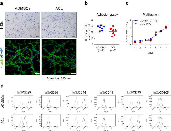
Fig. 1.
a) Morphology of anterior cruciate ligament (ACL)-derived cells and adipose-derived mesenchymal stem cells (ADMSCs) was observed by Giemsa staining and F-actin staining. Scale bar 200 µm. b) ACL-derived cells and ADMSCs were incubated in a growth medium for a day, and the number of cells was counted for an adhesion assay (n = 6). c) ACL-derived cells and ADMSCs were incubated in a growth medium for the number of days indicated, and the cell proliferation rate was evaluated using a WST assay (n = 3). d) CD29, CD34, CD44, CD45, CD90, or CD105 expression of cell surface was analyzed by fluorescence-activated cell sorting (FACS). Data are presented as the mean and standard deviation. DAPI, 4′,6-diamidino-2-phenylindole; H&E, haematoxylin and eosin; IgG, immunoglobulin G; N.S., not significant; OD, optical density.

