Figure 3.
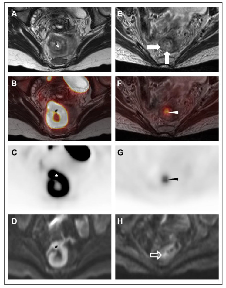
39-year-old woman with rectal adenocarcinoma (Subject #7) underwent FDG-PET/MRI at initial staging (A–D) and following TNT (E–H). Oblique axial high-resolution coronal T2-weighted images (A,E); oblique axial high-resolution coronal T2-weighted images with PET fusion (B,F), oblique axial attenuation-corrected PET images (C,G), and axial diffusion-weighted images (b = 1000 s/mm2) (D,H) are shown. At initial staging, a hypermetabolic, diffusion-restricting high rectal mass (*) was noted. There was no definite T3 disease, though assessment was limited by motion artifact and partial intussusception of the lesion. No suspicious lymph nodes were identified. Based on the limitations in T-stage determination, the patient underwent TNT. On restaging, both readers suspected some residual disease on MRI based on nodular T2-intermediate wall-thickening (closed arrows) and focal diffusion restriction (open arrow). Both readers assigned an mrTRG of 3. On the PET, there was a clear focus of hypermetabolism (arrowheads) in this same area, and both readers assigned a pmrTRG of 3. Although residual disease was already suspected based on the MRI alone, both readers reported a substantial increase in confidence based on the PET findings. Endoscopic biopsy confirmed residual disease, and the patient underwent resection with pathology showing ypT2 ypN0 disease.

