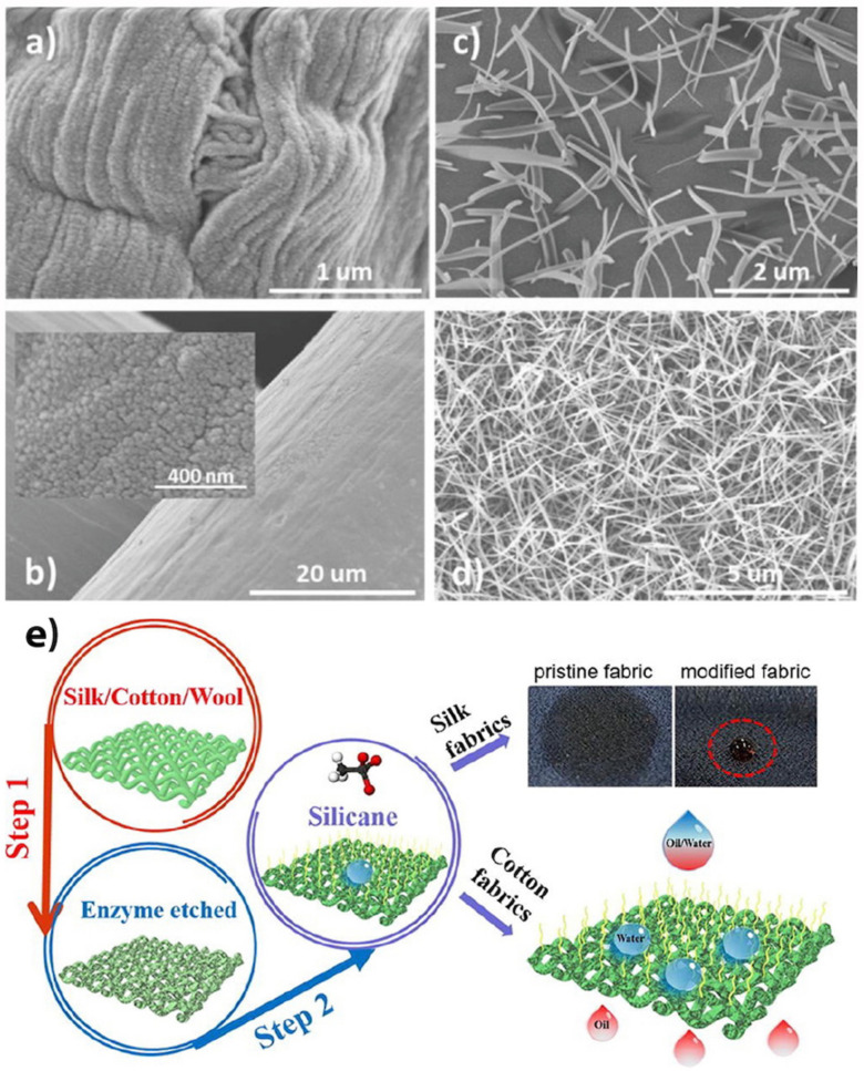
Figure 15.
SEM images of (a) cellulose filter, and (b) SS membrane after the first PECVD to form TiO2 nanofibers as the nucleation layer (step 1); (c,d) Top view SEM images of H2Pc nanowires created on TiO2 nucleation layer under the growth rate of <0.5 Ås−1 and >1.5 Ås−1, respectively. (Reproduced from [102] with permission from John Wiley and Sons). (e) The enzyme etched fabric is silanized via CVD to reduce the surface energy for oil/water separation application. (Reprinted from [105] with permission from Elsevier).

