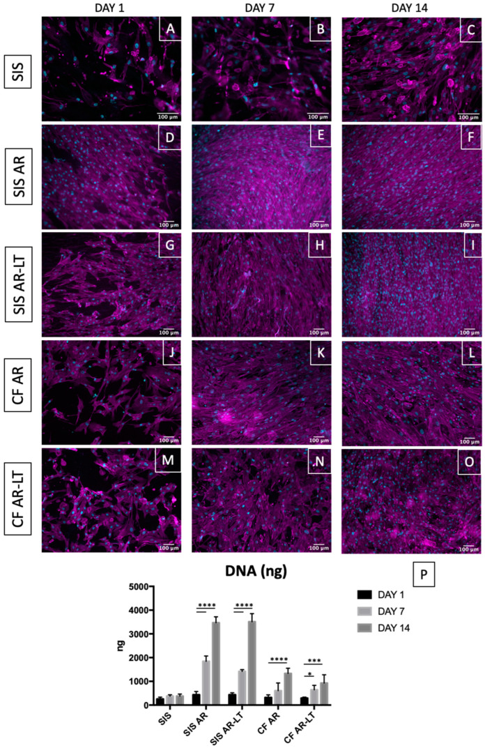
Figure 6.
Cell growth on decellularized tissue, hybrid membranes, and polycarbonate urethanes. Phalloidin was used to stain F-actin in magenta and DAPI to stain nuclei in cyan (A–O). Throughout the experimental period, a gradual cell increasing was observed in all groups, especially in the case of hybrid membranes and polymers alone (here, z-stacks are reported of epifluorescence images). DNA graph (n = 3) (P) shows a significant increase between days 1 and 7 and between days 1 and 14 in both SIS AR and SIS AR-LT; between days 1 and 14 in CF AR and days 1 and 7 and days 1 and 14 in CF AR-LT using Dunnett’s multiple comparison test (day 1 of each group was used as control group). No differences were found in the SIS group. * p < 0,05, *** p < 0.001 and **** p < 0.0001. Data show mean ± SD.

