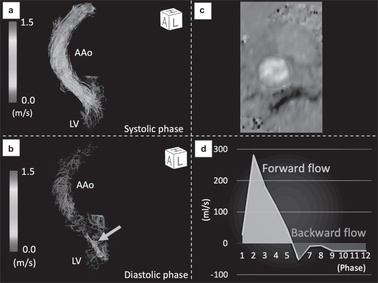
Fig. 1.
Quantitative flow measurements by 4D flow MRI in a female patient with AR. (a) Extreme helical or vortical patterns of forward flow were not observed in the systolic phase. (b) Regurgitation flow was clearly visualized with streamlined images in the diastolic phase (arrow). (c) By setting an arbitrary cross-section of a 3D image and drawing the region of interest, quantitative measurements in any cross-section can easily be performed. (d) Time-flow rate curve for one cardiac cycle of (12-phase). Forward flow was represented as integration of flow velocity curve in green and backward flow in red. AAo, ascending aorta; AR, aortic regurgitation; LV, left ventricle.

