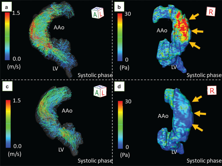
Fig. 2.
The change in wall shear stress before and after TAVI in a male patient with bicuspid aortic valve. Echocardiography revealed severe aortic stenosis with a transvalvular peak velocity of 5.3 m/s, mean gradient of 59 mmHg, and valve area of 0.83 cm2. 4D flow MRI demonstrated markedly accelerated flow (a) with extremely high wall shear stress (39.7 Pa) at the right wall of the ascending aorta (b, arrows). After TAVI, the helical flow decreased (c), and the wall shear stress decreased to 8.4 Pa (d, arrows). AAo, ascending aorta; LV, left ventricle; Pa, pascal; TAVI, transcatheter aortic valve implantation.

