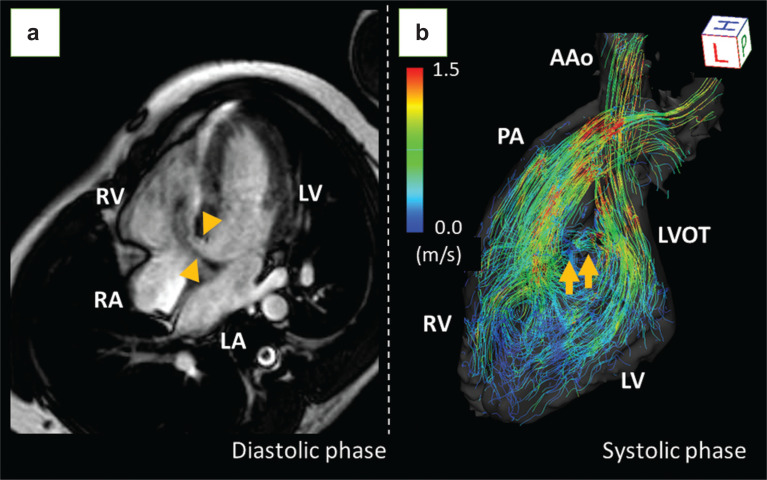
Fig. 3.
A case of VSD. Four-chamber cine MRI revealed a large VSD of 10 mm (a, arrowheads). The streamlined image of 4D flow MRI clearly depicts the flow jet of the VSD from the LVOT to the RV, as well as the laminar flow in the ascending aorta and the pulmonary artery (b, arrows). The pulmonary to systemic blood flow ratio (Qp:Qs) was 1.62:1. Although he had no symptoms, surgical repair was performed due to an elevated mean pulmonary arterial pressure of 25 mmHg with reduced right ventricular ejection fraction of 37%. AAo, ascending aorta; LA, left atrium; LV, left ventricle; LVOT, left ventricular outflow tract; PA, pulmonary artery; RA, right atrium; RV, right ventricle; VSD, ventricular septal defect.

