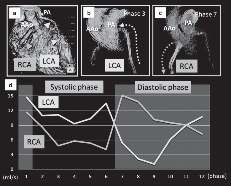
Fig. 4.
A case of Bland–White–Garland syndrome. (a) Computed tomography volume rendering image showing the RCA arising from the Valsalva sinus and the LCA arising from the PA (a). (b and c) 4D flow MRI clearly visualized the retrograde flow of the LCA (b) and the antegrade flow of the RCA (c) at different phases. (d) Quantitative assessments revealed that antegrade RCA flow was dominant in the diastolic phase, while retrograde LCA flow was dominant in the systolic phase. The flow volumes of both arteries were increased (LCA, 5.84 mL; RCA, 5.81 mL; stroke volume of the left ventricle, 61.7 mL), which suggested coronary steal phenomenon. AAo, ascending aorta; LCA, left coronary artery; PA, pulmonary artery; RCA, right coronary artery.

