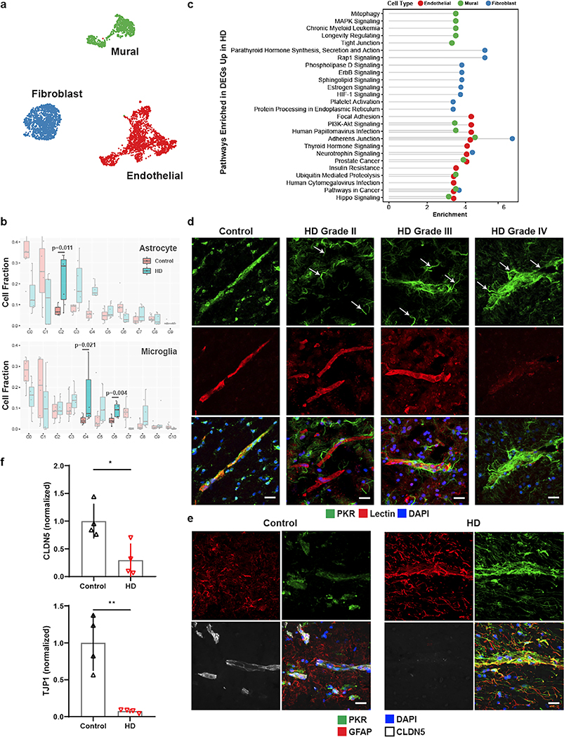
Figure 5. Innate immune activation related to cerebrovascular dysfunction in Huntington’s disease (HD).
a. UMAP of integrated cerebrovasculature cells in post mortem control and HD human patients. b. Cell fraction analysis of astrocyte and microglia subclusters; statistics shown for highlighted clusters using the Wilcoxon rank-sum test. Center-line denotes the median; box limits denote the upper and lower quartiles; and whiskers denote the 1.5x interquartile range. c. Pathway analysis of the top 10 enriched upregulated pathways in HD endothelial, mural, and fibroblasts cells. d. PKR immunostaining in perivascular glial processes across various HD grades. PKR is normally detected in the vasculature in control samples; arrows indicate PKR-immunopositive vasculature-coupled glial processes that become apparent in HD samples only. e. PKR immunostaining co-localizes with GFAP and engulfs blood vessels with low CLDN5 expression. f. Western blot quantification for tight junction proteins CLDN5 and TJP1. Scale bar, 20μm. WB: two-tailed t test, *p < 0.01, **p < 0.001. Error bars denote standard deviation.

