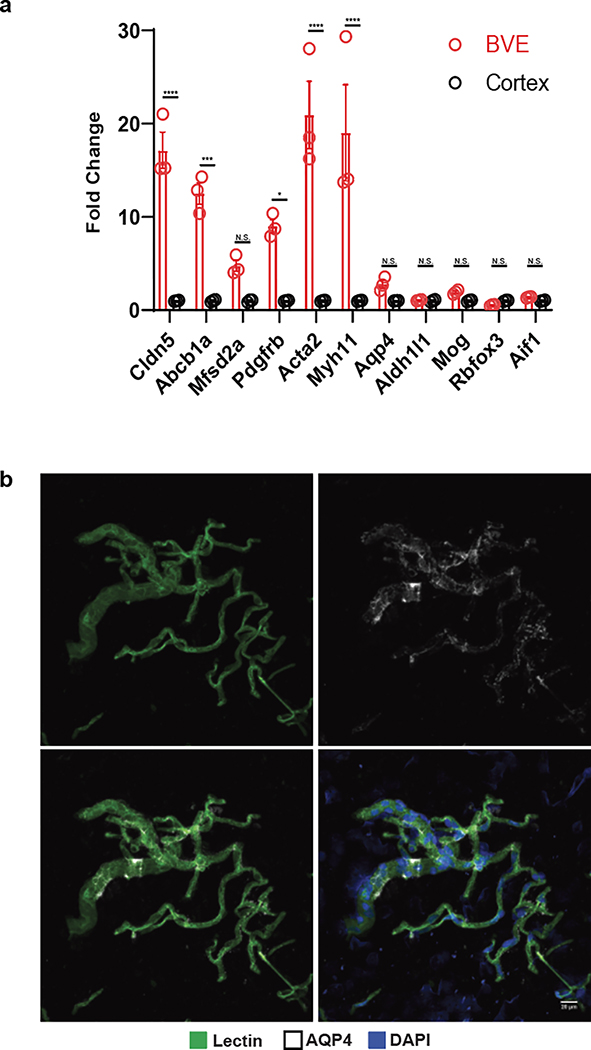
Extended Data Figure 1. Validation of Blood Vessel Enrichment (BVE) protocol.
a. qPCR of canonical cell type markers for endothelial (Cldn5, Abcb1a, Mfsd2a), mural (Pdgfrb, Acta2, Myh11), astrocytes (Aqp4, Aldh1l1), oligodendrocytes (Mog), neurons (Rbfox3), and microglia (Aif1) from mouse cortex, n=3, two-tailed t test, *p < 0.01, **p < 0.001, ***p < 0.0001,****p < 0.00001, n.s. = not significant. b. Immunofluorescence of blood vessels enriched from mouse cortex using the BVE protocol. Scale bar, 20μm

