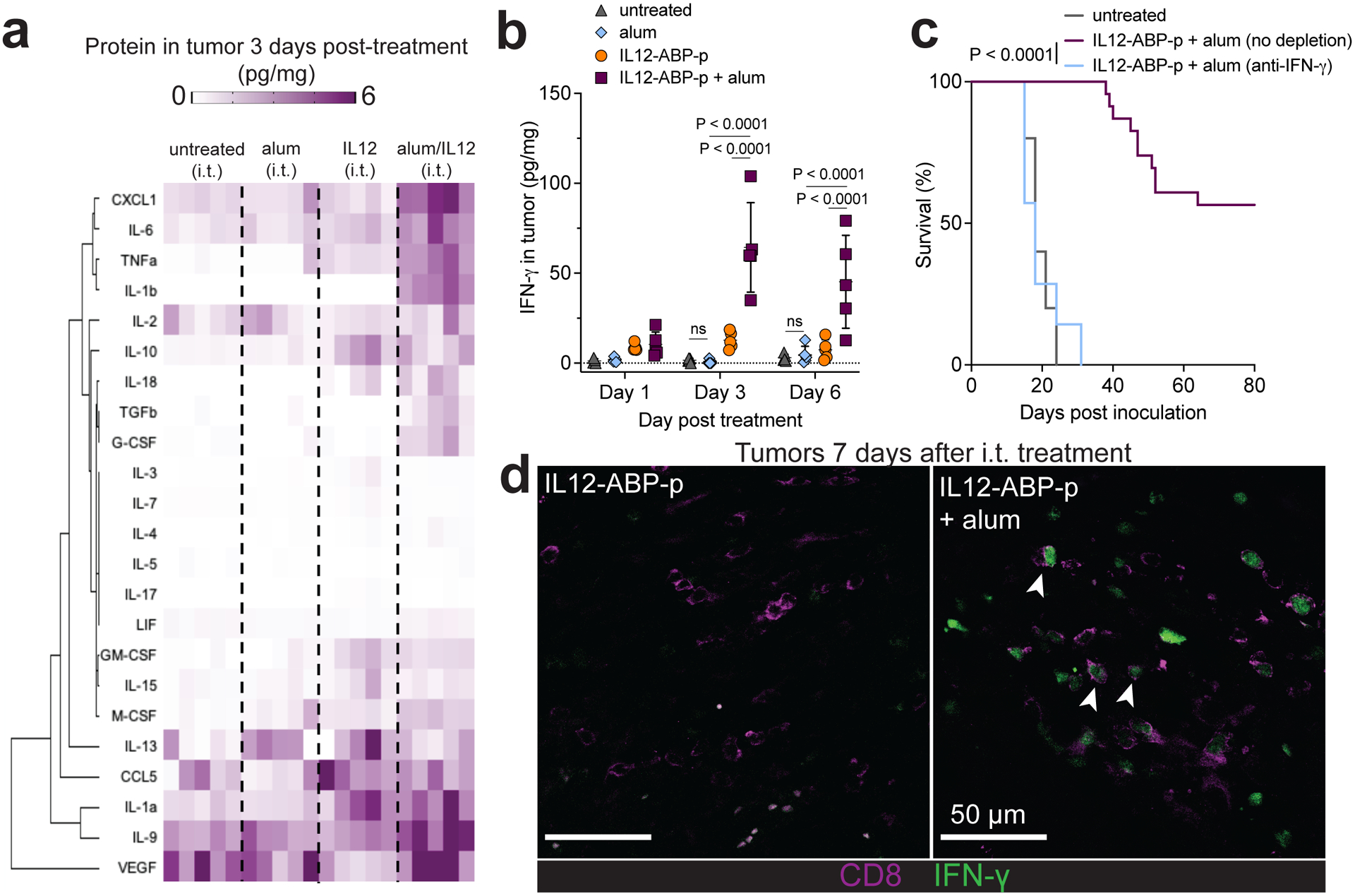
Fig. 6 |. IFN-γ production is critical for alum-anchored IL-12-mediated tumour rejection.

a-b, Mice were inoculated s.c. in the flank with 106 B16F10 tumour cells and treated on day 6 with no i.t. treatment (n=5 mice per group), alum (100 ug) i.t., IL-12-ABP-p (20 ug) i.t. or IL-12-ABP-p (20 ug) + alum (100 ug) i.t. combined with systemic anti-PD1 (200 ug per dose) on days 6 and 9. Tumours were isolated and cytokine/chemokine levels were quantified by Luminex 3 days post treatment (a) and IFN-γ amounts in tumours 1, 3 or 6 days after treatment were quantified by ELISA (mean ± SD, b). c, Overall survival for mice bearing B16F10 tumours and treated as in a in the presence or absence of anti-IFN-γ starting 1d before i.t. treatment and every 2d thereafter (untreated n=10 mice/group, treated n=23 mice/group, and treated + anti-IFN-γ n=7 mice/group). d, IFN-γ reporter mice bearing B16F10 tumours were treated as in a with AF405-labeled IL-12-ABP-p alone or combined with alum. Shown are representative histological tumour cross sections 7 days after treatment with IFN-γ in green and CD8 in magenta (n = 3 mice/group; scale bars, 50 um, white arrows point to IFN-γ+CD8+ T cells). ABP refers specifically to ABP10. P values were determined by the log-rank (Mantel-Cox) test (c) and two-way (b) ANOVA followed by Tukey’s multiple comparison test using GraphPad PRISM.
