
FIG. 1.
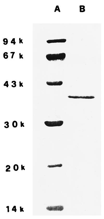
Electrophoretic pattern of the proteins present in a purified sample of P. aeruginosa porin (lane B). The SDS-polyacrylamide gel (12%) was stained with Coomassie blue. The molecular mass markers (daltons) are in lane A.
Official websites use .gov
A
.gov website belongs to an official
government organization in the United States.
Secure .gov websites use HTTPS
A lock (
) or https:// means you've safely
connected to the .gov website. Share sensitive
information only on official, secure websites.
Electrophoretic pattern of the proteins present in a purified sample of P. aeruginosa porin (lane B). The SDS-polyacrylamide gel (12%) was stained with Coomassie blue. The molecular mass markers (daltons) are in lane A.