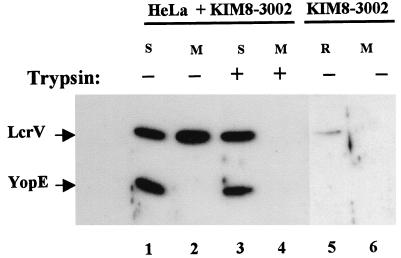
FIG. 1.
LcrV partitions to the cell-free medium and HeLa soluble fractions. Duplicate wells containing HeLa monolayers were infected with Y. pestis KIM8-3002 at an MOI of 10 (lanes 1 to 4). As a control, an equal dose of Y. pestis KIM8-3002 was also added to a well lacking HeLa cells (lanes 5 and 6). After incubation at 37°C-5% CO2 for 4 h, trypsin was added to 100 μg/ml to one replicate of infected HeLa cells. Yersinia-infected HeLa cultures were fractionated, and the cell-free medium (M) and water-lysate soluble (S) fractions were further analyzed. Cell-free medium (M; lane 6) from the Yersinia-only well corresponded to the culture supernatant after removal of bacteria by centrifugation. The bacteria were then treated with cold water and pelleted, and the resulting supernatant corresponding to the water-released (R; lane 5) fraction was removed. Samples from each fraction representing 0.3% of the original culture were resolved in a 12% polyacrylamide gel. LcrV and YopE were detected by probing immunoblots with α-HTV and α-YopE followed by secondary antibodies conjugated to horseradish peroxidase and development with ECL reagent.

