. 2022 Nov 8;11:e82709. doi: 10.7554/eLife.82709

Figure 5. Loss of sloth1-sloth2 causes neurodegeneration.

(A–C) Transmission electron microscopy (TEM) images of sectioned adult eye photoreceptors (left) and quantification of photoreceptor number and aberrant photoreceptors (right). Scalebar is 2 µm. Filled red arrows indicate dead or dying photoreceptors. Open red arrows indicate unhealthy photoreceptors. Error bars show mean with SD. N≥8 ommatidium per genotype. (A) Four weeks old raised in a 12 hr light/dark cycle. (B) 3 days old raised in a 12 hr light/dark cycle. (C) Four weeks old raised in 24 hr dark. (D) Confocal microscopy of adult eye photoreceptors stained with phalloidin (green) and anti-Rh1 (red). Animals were 4 weeks old and raised in a 12 hr light/dark cycle. Arrowheads indicate photoreceptors with higher levels of Rh1.

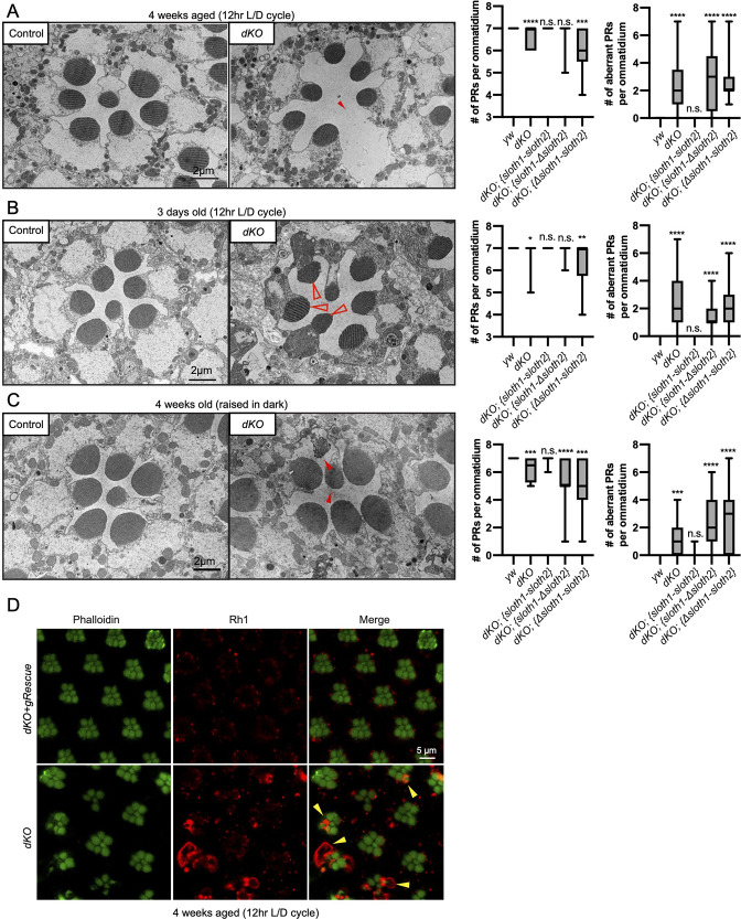
Figure 5.

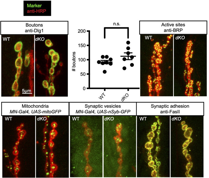
Figure 5—figure supplement 1. Related to Figure 5.

Figure 5—figure supplement 1.

Confocal microscopy images of 3rd instar larval NMJ at muscle 6/7 segment A2. Antibodies or fluorescent proteins (green) mark synaptic components and anti-HRP (red) marks neurons. Comparison of wild-type to dKO. Graph shows quantification of synaptic bouton number by anti-Dlg1 staining. Significance of dKO bouton number was calculated with a T-test compared to WT. Error bars show mean with SD. N≥7 NMJs (each from a different animal).
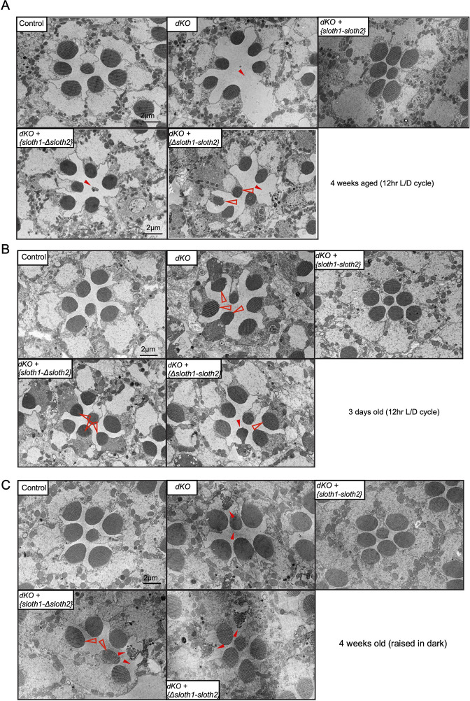
Figure 5—figure supplement 2. Related to Figure 5.

Figure 5—figure supplement 2.

(A–C) Transmission electron microscopy (TEM) images of sectioned adult eye photoreceptors from indicated genetic backgrounds with accompanying quantification of photoreceptor number and aberrant photoreceptors. Scalebar is 2 µm. Filled red arrows indicate dead or dying photoreceptors. Open red arrows indicate unhealthy photoreceptors. Error bars show mean with SD. (A) Animals were 4 weeks old and raised in a 12 hr light/dark cycle. (B) Animals were 1–3 days old and raised in a 12 hr light/dark cycle. (C) Animals were 4 weeks old and raised in the dark.
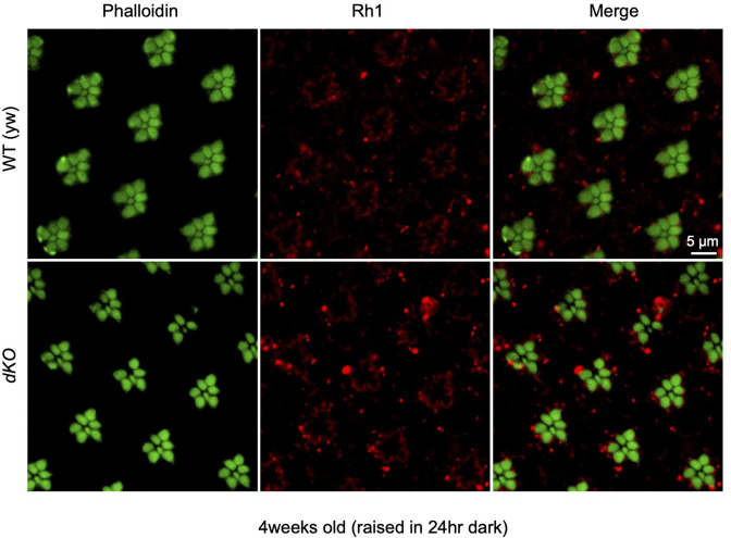
Figure 5—figure supplement 3. Related to Figure 5.

Figure 5—figure supplement 3.

Confocal microscopy of adult eye photoreceptors stained with phalloidin (green) and anti-Rh1 (red). Animals were 4 weeks old and raised in the dark. Arrows indicate photoreceptors with higher levels of Rh1.