. 2022 Nov 8;11:e82709. doi: 10.7554/eLife.82709

Figure 6. Sloth1 and Sloth2 localize to mitochondria.

(A) Confocal microscopy of S2R+ cells transfected with Sloth1-FLAG or Sloth2-FLAG and stained with anti-FLAG (green) and anti-ATP5alpha (red). DAPI (blue) stains nuclei. (B–D) SDS-PAGE and western blotting of S2R+ cellular fractions. WCL = Whole Cell Lysate, cyto.=cytoplasmic lysate, mito.=mitochondrial lysate. Mitochondrial control = ATP5alpha, cytoplasmic control = alpha-tubulin. Each lane loaded equal amounts of protein (15 µg/lane). Blots were stripped and reprobed after detection of each antigen. (B) Transfected Sloth1-FLAG or Sloth2-FLAG. (C) Stable cells expressing copper-inducible Sloth1-SBP or Sloth2-SBP. (D) Stable cells expressing copper-inducible Sloth1-SBP or Sloth2-SBP.

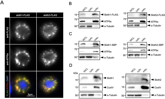
Figure 6.

Figure 6—figure supplement 1. Related to Figure 6.

Figure 6—figure supplement 1.

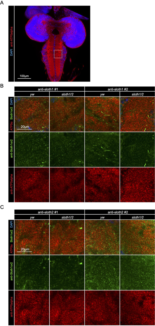
Confocal microscopy of 3rd instar larval brain with antibody staining. Anti-Sloth1 or Anti-Sloth2 (green), mitochondria labeled with anti-ATP5alpha (red), and nuclei labeled with DAPI (blue). Wild-type (yw) or sloth1/2 KO. (A) Zoom out of entire brain showing region imaged in panels B and C. Scale bar 100 µm. (B) Results using two independent anti-Sloth1 antibodies (#1 and #2). Scale bar 20 µm. (C) Results using two independent anti-Sloth2 antibodies (#1 and #2). Scale bar 20 µm.
Figure 6—figure supplement 2. Related to Figure 6.

Figure 6—figure supplement 2.

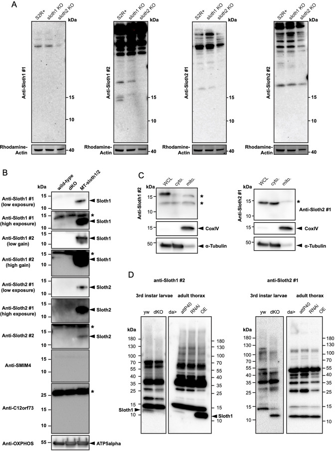
SDS-PAGE and western blotting using anti-Sloth1 and anti-Sloth2 antibodies of cell and mitochondrial lysates. Two independent (#1 and #2) anti-Sloth1 and Anti-Sloth2 antibodies were tested. Arrowheads indicated expected band, asterisks indicate unrelated band(s). Tricine gels were used. (A) S2R+ whole cell lysates isolated from indicated genotypes. Rhodamine-Actin used as loading control. (B) S2R+ mitochondrial lysates isolated from indicated genotypes. Anti-ATP5alpha used as loading control. Mitochondrial control = ATP5alpha, cytoplasmic control = alpha-tubulin. (C) Mitochondrial fractions isolated from wild-type S2R+ cells. WCL = Whole Cell Lysate, cyto.=cytoplasmic lysate, mito.=mitochondrial lysate. Blots were stripped and reprobed after detection of each antigen. (D) Mitochondrial lysates isolated from 3rd instar larvae or adult thorax mitochondrial isolation of indicated genotypes. ‘da>’ indicates da-Gal4 crossed with attP40 (wild-type), RNAi (UAS-shRNA-sloth1/2), OE (UAS-sloth1/2 transcript).