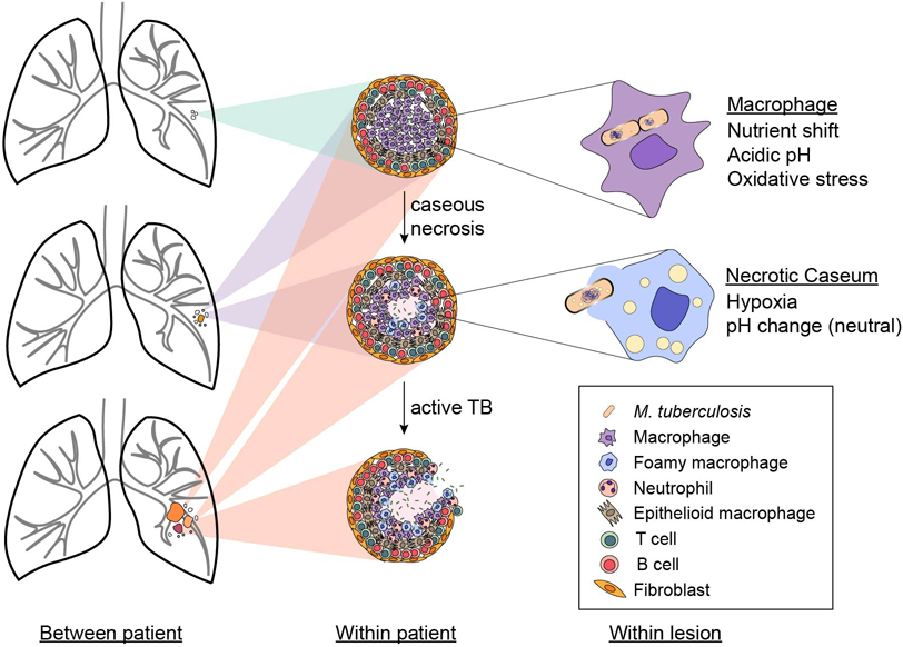
Figure 2. Environmental heterogeneity.
Microenvironments in tuberculosis lesions (granulomas) vary among patients, within one patient, and even within a lesion as shown with examples in the first, second, and third columns, respectively. Lesion composition is dependent on the stage of disease progression and the network of immune cells present. Many types of cells respond to infection with Mycobacterium tuberculosis, including macrophages, neutrophils, T and B cells, epithelioid cells, and fibroblasts, and populate cellular granulomas. M. tuberculosis in infected macrophages encounter an environment characterized by low pH, oxidative stress and abundant lipids. As M. tuberculosis utilizes these host lipids, it secretes lipid vesicles into the macrophage cytoplasm, inducing some of the infected macrophages to become foamy with lipid droplets. Cytoplasmic debris from necrotic macrophages form the granuloma's caseous center, which may be acidic or neutral and vary in oxygen content.

