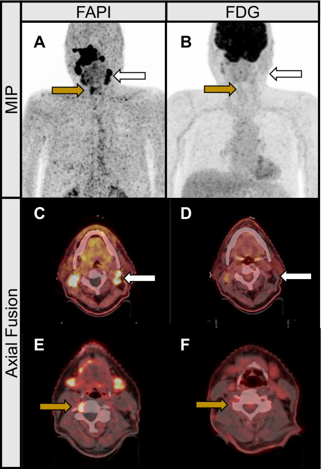
Fig. 2.

Cervical nodal metastasis with high FAPI uptake and low FDG uptake maximum intensity projections (A, B) and axial fusion images of a 73-year-old male with nasopharyngeal cancer prior to radiation therapy. The patient was upstaged with newly emerged nodal metastasis on both sides of the neck, noticed only in the FAP scans (C, D; white arrows). [.68 Ga]-FAPI PET/CT also revealed one cervical lymph node metastasis in close proximity to the cervical spinal cord (at C4 level) that would have been missed in the FDG-scans (E, F; yellow arrows).
