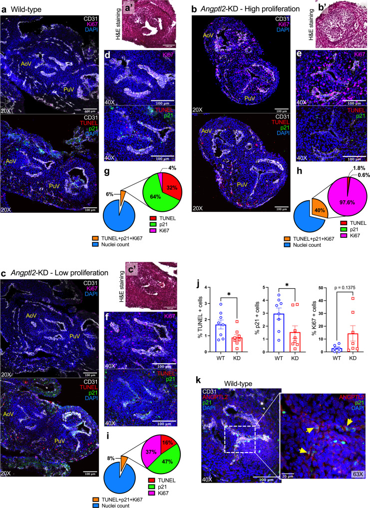
Fig. 3. AoV defects in Angptl2-KD mouse embryos are associated with an impairment in the balance between embryonic apoptosis, senescence and proliferation.
Representative images (of n = 3 independent experiments) of TUNEL assay and Ki67, p21 and CD31 protein expression by immunofluorescence in cardiac sections from E14.5 embryos from WT (a) and Angptl2-KD mice (b, example of High proliferation; c example of Low proliferation), showing both aortic valve (AoV) and pulmonary valve (PuV). In the same WT or Angptl2-KD embryo AoV section, H&E staining (a’–b’–c’) and its corresponding TUNEL assay, Ki67 and p21 protein expression (d–f) are shown. Pie charts illustrate the percentage of marked cells of the total nuclei, and the corresponding proportion (%) of TUNEL, p21 and Ki67-positive cells within the marked cells (g–i). j For TUNEL/p21: The graphs represent means ± SEM of n = 4 embryos per genotype (2 sections analyzed/embryo; *: p < 0.05 determined with unpaired t-test); For Ki67: The graph represents means ± SEM of n = 6 embryos for WT and n = 7 embryos for Angptl2-KD (1 section analyzed/embryo; p = 0.1375 determined with Mann–Whitney U test). k Representative images (of n = 3 independent experiments) of ANGPTL2 and p21 protein expression by immunofluorescence in AoV from E14.5 embryos from WT mice. Higher magnification (×63) shows cells co-expressing ANGPTL2 and p21 (indicated by yellow arrows). DAPI staining was used to visualize cell nuclei.

