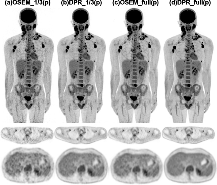
Fig. 4.
PET images of a 31-year-old male patient diagnosed with Hodgkin's lymphoma in the prospective study. After injection of 85 MBq 18F-FDG, the patient underwent a PET scan with 6 min per bed position 77 min post-injection. To comprehensively compare the clinical images reconstructed by the two algorithms, we further reconstructed the data with 1/3 and full of the acquisition duration for OSEM and DPR algorithms. Both the MIP (upper row) and transverse images (middle row) demonstrated improved lesion detectability of the DPR images compared to the OSEM images with the same acquisition duration (a–d). The transverse image in the liver plane showed reduced image noise when comparing subfigures a–d, while the image noise in the subfigures b and c was at a comparable level

