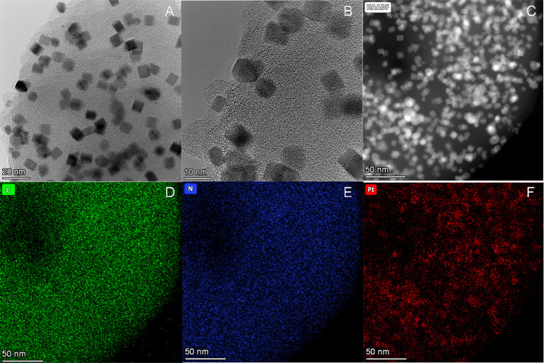
Figure 2.
Structural and morphological characterization of Purp@COP. (A) Transmission electron microscope (TEM) images of Purp@COP (scale, 20 nm). (B) High-resolution TEM images of Purp@COP (scale, 10 nm). (C) High-angle annular dark-field scanning transmission electron microscope (HAADF-STEM) of Purp@COP (scale, 50 nm). (D–F) Energy-dispersive spectrum (EDS) elemental mapping images of C, N, and Pt (scale, 50 nm).

