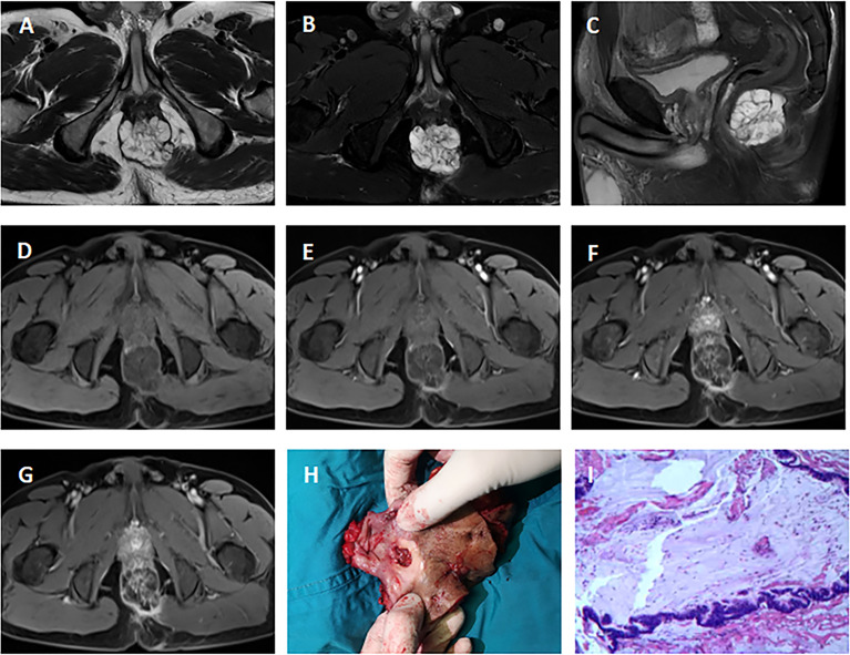
Figure 1.
MRI of a 51-year-old man with mucinous adenocarcinoma. (A): Axial T2-weighted image; (B): Axial fat-suppressed T2-weighted image; (C): Sagittal fat-suppressed T2-weighted image; (D–G): Axial dynamic contrast-enhanced images. The tumor grew across the midline. This lesion was multiloculated, cauliflower-like, markedly hyperintense on fat-suppressed T2-weighted image. Axial dynamic contrast-enhanced images showed progressive heterogeneous enhancement, and the boundary of the enhanced region was clear. (H): Clinical photograph showed the abdominoperineal resection specimen (the proximal intestinal canal was cut up). (I): The tumor was rich in mucus (hematoxylin and eosin staining; original magnification, ×100).

