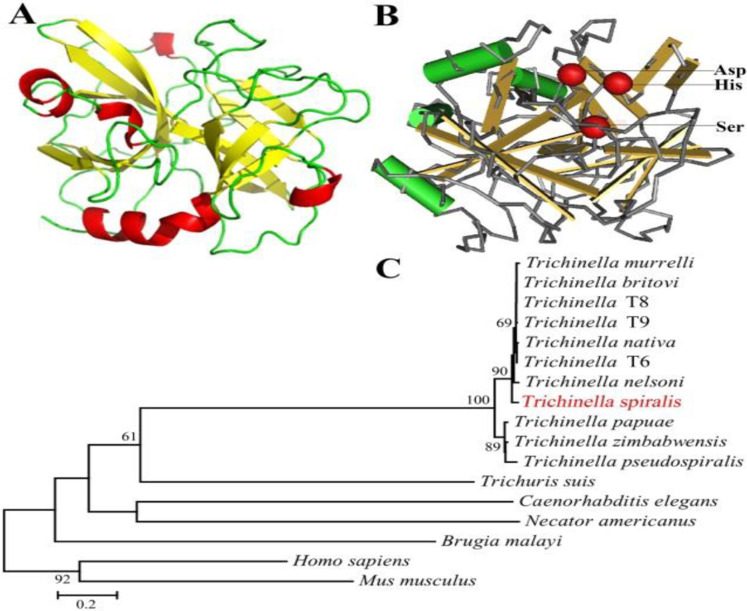
Fig. 2:
Predicted three-dimensional model of TsSP1.1 and phylogenetic trees of serine proteases of 17 organisms. A: Predicted three-dimensional structure of TsSP1.1, there are 5 α-helixes (in red), 11 β-strand (in yellow), and 15 irregular coils (in green). B: Three serine protease-specific enzyme active sites (Asp, Ser and His) of TsSP1.1 are marked red. C: Phylogenetic tree of serine proteases from 17 organisms calculated using MEGA with NJ method

