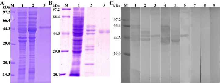
Fig. 3:
Identification of rTsSP1.1. A: SDS-PAGE analysis of rTsSP1.1. Lane M: Protein marker; Lane 1: ly-sate of recombinant pQE-80L/TsSP1.1 prior to induction; Lane 2: lysate of recombinant pQE-80L/TsSP1.1 following induction; Lane 3: purified rTsSP1.1. B: SDS-PAGE analysis of muscle larva (ML) somatic crude antigens, ML excretion/secretion (ES) antigens and rTsSP1.1. Lane M: protein marker; Lane 1: ML crude antigens; Lane 2: ML ES antigens; Lane 3: rTsSP1.1. C: Western blotting of rTsSP1.1. ML crude antigens (lane 1), ML ES antigens (lane 2), and rTsSP1.1 (lane 3) were recognized by infected sera. Native TsSP1.1 in ML crude antigens (lane 4), ML ES antigens (lane 5) and rTsSP1.1 (lane 6) were identified by anti-rTsSP1.1 serum; but native TsSP1.1 in ML crude antigens (lane 7), ML ES antigens (lane 8) and rTsSP1.1 (lane 9) were not recognized by normal serum

