Figure 1.
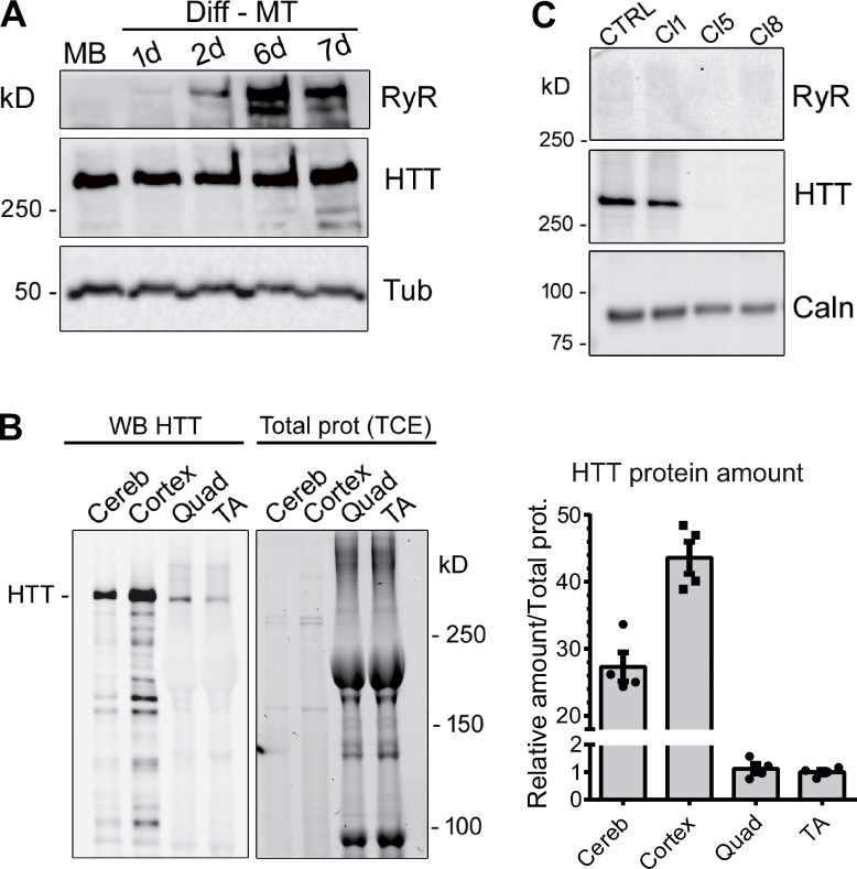
Western blot analysis of HTT protein. (A) Western blot analysis of immortalized human control myoblasts (MB) and after 1, 2, 6, and 7 d in differentiation medium (Diff-MT). The HTT protein is stably expressed in myoblasts or myotubes. The RyR1 protein expression level increases during differentiation. Tubulin is used as the loading control. (B) 5 µg mouse brain (cerebellum or cortex) was analyzed by Western blot, together with 50 µg mouse skeletal muscle (quadriceps, Quad; tibialis anterior, TA). HTT protein amounts (right) were normalized to the total protein amounts determined with TCE. Data are presented as mean ± SEM of four mice. (C) Western blot analysis of HTT amount in human immortalized myoblasts CTRL and clone 1-CRTL (Cl1), and in HTT-KO clone 5 (Cl5) and 8 (Cl8). Calnexin is used as the loading control. Source data are available for this figure: SourceData F1.

