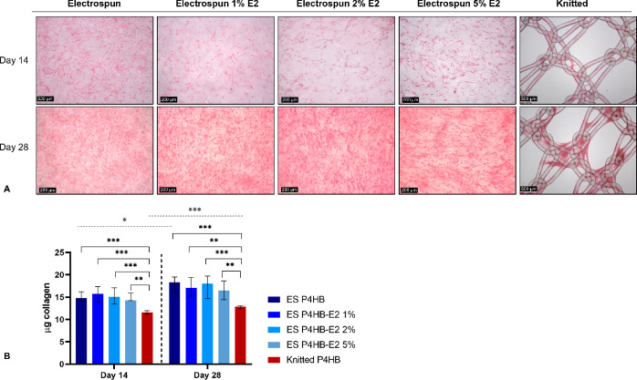
Figure 4.
Collagen deposition. Collagen deposition was imaged after a Picrosirius Red stain on days 14 and 28. (A) Representative images demonstrate increased collagen deposition over time and collagen was more evenly distributed over the electrospun (ES) P4HB scaffolds as compared to the knitted P4HB. (B) Collagen deposition increased over time, which reached significant differences for ES P4HB and knitted P4HB (gray dashed line above figure). Collagen deposition was significantly higher for all ES P4HB as compared to knitted P4HB on both days 14 and 28 (black brackets). Data is reported as median with IQR. *p < 0.05, **p < 0.01, ***p < 0.001.

