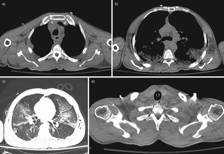
FIGURE 4.
Initial non-contrast computed tomography (CT) of the chest and neck; case two. a) Axial CT image through the upper thorax shows fluid collections in the anterior mediastinum and posterior to the oesophagus. b) Axial CT chest image at the level of the carina shows an enlarged pre-carinal lymph node with bilateral air space opacities posteriorly and extrathoracic soft tissue gas along the chest wall. c) Axial CT image through the mid-thorax with lung windows demonstrates bilateral airspace opacities and a small right pneumothorax but no visible mediastinal gas. A chest tube is visible in the right hemithorax. d) Axial CT image through the lower neck shows no cervical abscess or suspicious gas collections.

