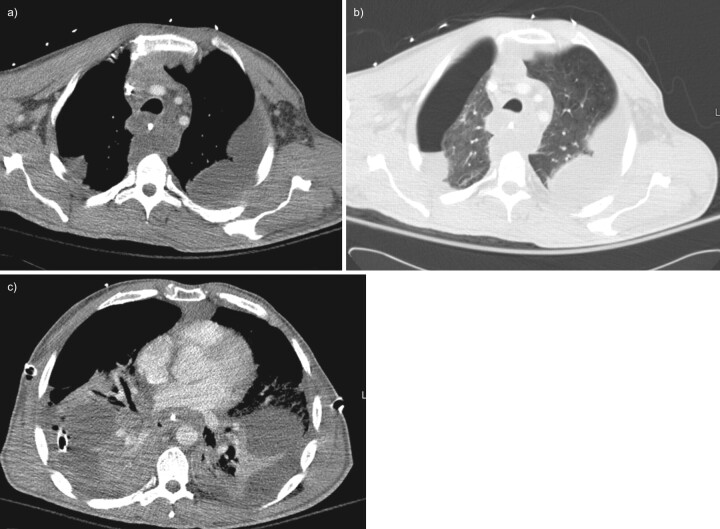
FIGURE 6.
Contrast-enhanced computed tomography (CT) of the chest; case two, 4 days after presentation. a) Axial CT image of the upper thorax at the level of the great vessels on mediastinal windows shows fluid attenuation within the mediastinal fat and multiple bilateral loculated pleural fluid collections. b) Axial CT image of the upper thorax at the level of the great vessels on lung windows shows a right hydropneumothorax. c) Axial CT image of the mid-thorax on mediastinal windows shows loculated pleural fluid collections bilaterally with bilateral chest tubes.

