
Figure 4.
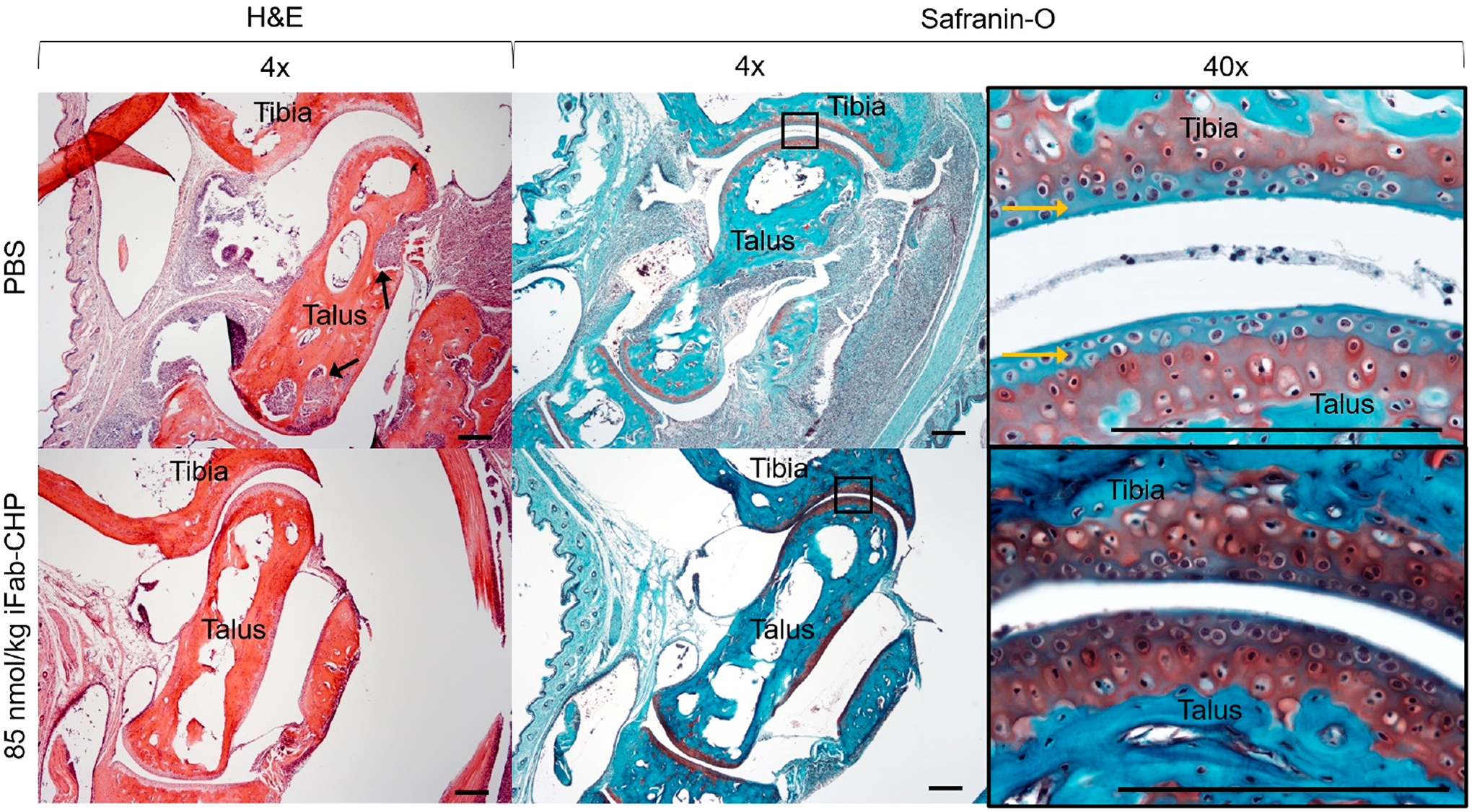
Histological staining of hTNFα transgenic mice tibia-talus joints with either hematoxylin and eosin (H&E) (first column) or Safranin-O (red) with Fast Green (blue) (second and third columns). Mice were injected at disease onset with either 85 nmol/kg iFab-CHP or PBS, with an additional injection 7 weeks post-initial injection, and sacrificed at 10 weeks post-initial injection. H&E staining revealed a greater degree of synovial hyperplasia and inflammatory cell infiltration (black arrows) in PBS treated mice. Additionally, Saf-O/Fast Green staining revealed more intact hyaline articular cartilage (sulfated proteoglycans stained red) in iFab-CHP vs PBS treated mice. Yellow arrows point to negative hyaline cartilage staining. Scale bars = 200 μm.