Fig. 2.
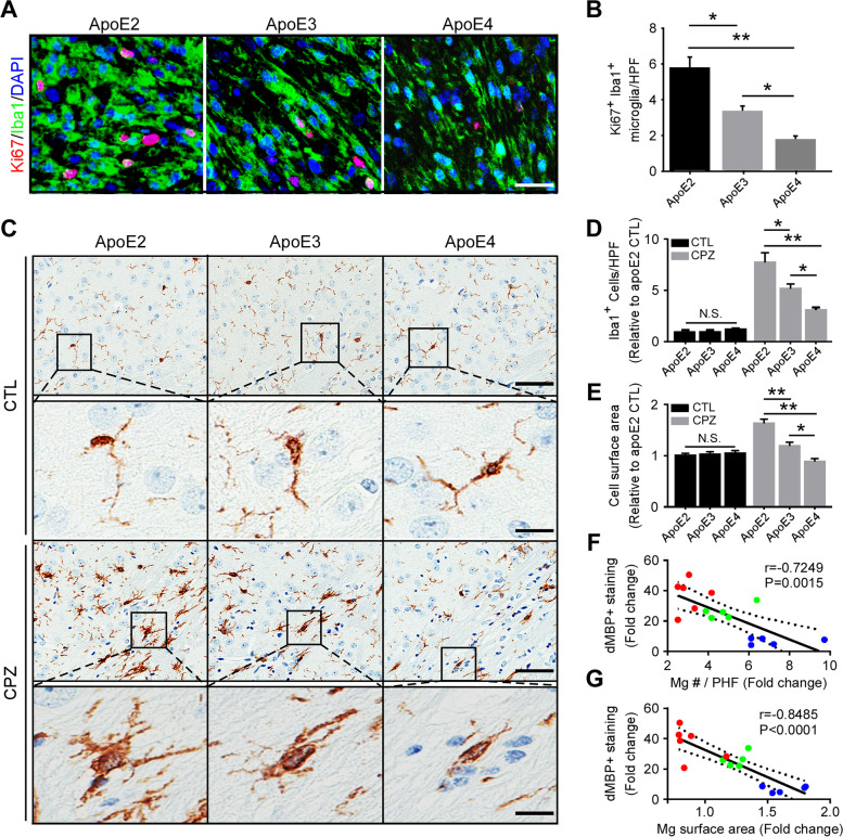
ApoE isoform-dependent effects on microglial proliferation and morphological changes. A Brain samples from CPZ-treated apoE-TR mice (n = 12–13/genotype) were subjected to immunofluorescence staining with Ki67 (for proliferation) and Iba1 (for microglia). Representative images of Ki67+Iba1+ microglia upon CPZ-induced demyelination are shown. Scale bar, 15 µm. B The number of Ki67+Iba1+ microglia per high-power field (HPF) was quantified. C Representative images of Iba1+ microglia in the CC area are shown. Scale bar, 25 µm (low magnification); 10 µm (high magnification). D The numbers of Iba1+ microglia per high-power field (HPF; 3 HPFs/mouse) were analyzed. E The surface area of Iba1+ microglia in the CC area were quantified. F A negative correlation was observed between the fold change of microglia number (Mg #) and the fold change of dMBP+ staining upon CPZ treatment. G A negative correlation was shown between the fold change of microglial surface area and the fold change of dMBP+ staining upon CPZ treatment. Blue dots, apoE2; Green dots, apoE3; Red dots, apoE4. The Pearson correlation coefficient (r) and P values are shown. Values are mean ± SEM. Two-way ANOVA. *P < 0.05; **P < 0.01. N.S., not significant

