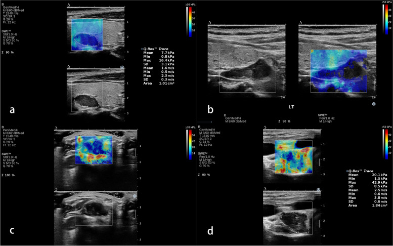
Fig. 1.
Four qualitative shear wave elastography patterns of parathyroid lesions. a The “negative” pattern was defined as no obvious color difference around and inside the lesion, displaying a homogeneously blue pattern; b The “void center” pattern was defined as the absence of color filling in the center of the lesion; c The “stiff rim” pattern was defined as increased stiffness (coded in orange or red) in the peritumoral region as compared with the stiffness in the surrounding soft tissues and the interior lesion tissues; d The “colored lesion” pattern was defined as the heterogeneously intralesional multicolor appearance

