Fig. 2.
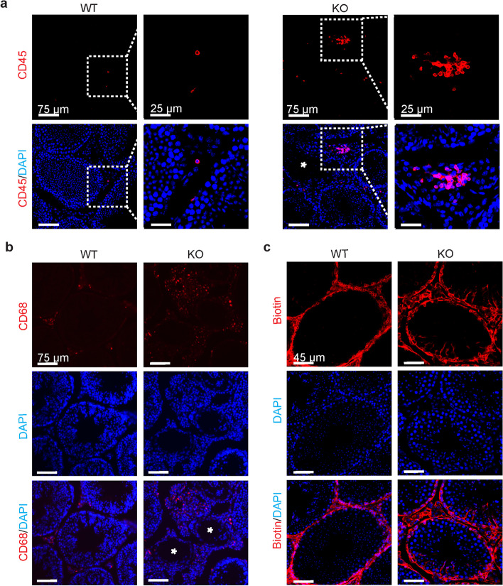
Comparison of the integrity of the blood–testis barrier between WT and Alkbh5-deficient mice. a, b Immunofluorescence analysis showing CD45+ (a) and CD68+ (b) cells infiltrated into the stroma in the testis of wild-type (WT) (n = 3) and Alkbh5-deficient (KO) (n = 3) mice, respectively (asterisk indicates morphologically damaged seminiferous tubule). c BTB assay detecting the integrity of the blood–testis barrier in WT (n = 3) and Alkbh5-KO mice (n = 3)

