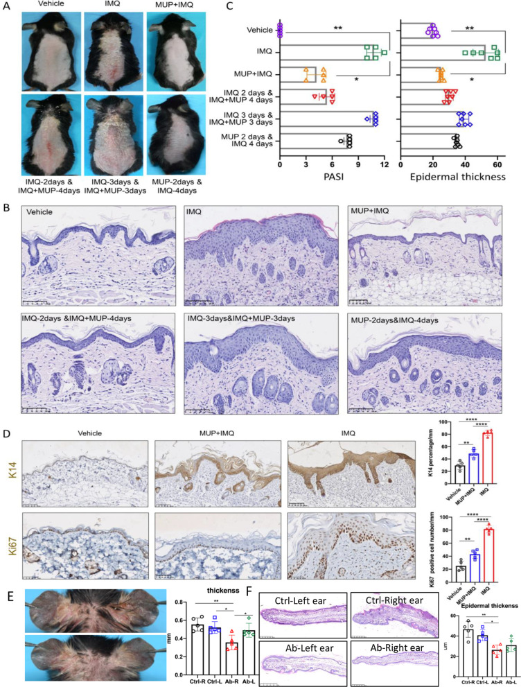
Fig. 2.
Mupirocin administration attenuated IMQ-treated mice. A Representative clinical presentations of vehicle-treated mice, IMQ-treated mice and MUP + IMQ-treated mice (top line from left to right), IMQ-2 days & IMQ + MUP-4 days-treated mice, IMQ-3 days & IMQ + MUP-3 days-treated mice and MUP-2 days & IMQ-4 days-treated mice (bottom line from left to right). B Representative H&E images of dorsal skin from different six treated mice described as (A). Scale bar = 100 μm. C PASI score (left) and epidermal thickness (right) was performed of day 6 from the six groups described as (A) (n = 5 ± SEM). *P < 0.05, **P < 0.01 vs. IMQ. D Representatives IHC images and quantification analysis of keratin14 (top) and Ki67 (bottom). Scale bar = 100 μm. (n = 5 ± SEM). ** P < 0.01 vs. vehicle, ****P < 0.0001 vs. IMQ). E Representative clinical presentations of IMQ-treated mice of the 0.9% NaCl-injection group (right ear) (top) and anti-IARS-injection group (10ul, abcam,ab315333, from 2nd to 5th day) (right ear) (bottom). Quantification analysis of ear thickness of the 0.9% NaCl-injection group and anti-IARS-injection group. (n = 5 ± SEM). * P < 0.05** P < 0.01. F Representative H&E images(left) and the average epidermal thickness(right) of ear skin from IMQ-treated different four mice groups described as (E). Scale bar = 100 μm. (n = 5 ± SEM). *P < 0.05, **P < 0.01

