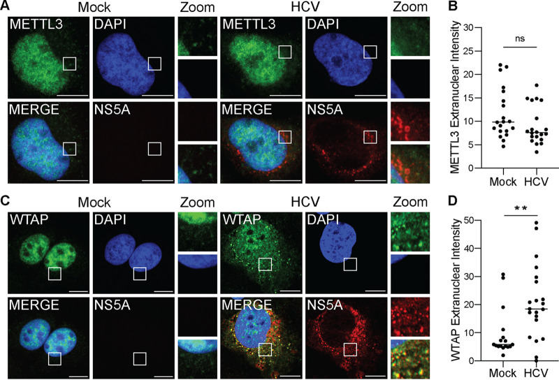
FIG 1.
HCV infection alters the subcellular localization of the m6A accessory protein WTAP. (A and C) Confocal micrographs of mock- or HCV (48 h, MOI of 0.3)-infected Huh7 cells stained with DAPI and antibodies against HCV NS5A and either METTL3 (A) or WTAP (C). Zoom is taken from area in the white box. (B and D) Quantification of the fluorescent signal intensity in the extranuclear region of Huh7 cells for METTL3 (B) or WTAP (D), as described in Materials and Methods for fields of mock (NS5A-negative)- or HCV-infected (NS5A-positive) cells. Scale bars = 10 μM. Graph shows mean ± SD; n = 21 fields. Data were analyzed by Welch’s unequal variances t test (**, P < 0.01; ns, not significant).

